Muscle function impairment in cancer patients in pre-cachexia stage
All claims expressed in this article are solely those of the authors and do not necessarily represent those of their affiliated organizations, or those of the publisher, the editors and the reviewers. Any product that may be evaluated in this article or claim that may be made by its manufacturer is not guaranteed or endorsed by the publisher.
Published: 22 June 2020
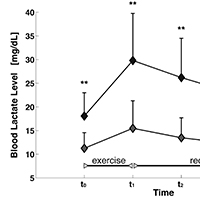
Cancer cachexia has been reported to be directly responsible for at least 20% of cancer deaths. Management of muscle wasting in cancer-associated cachexia appears to be of pivotal importance for survival of patients. In this regard, it would be interesting to identify before its patent appearance eventual functional markers of muscle damage, to plan specific exercise protocols to counteract cachexia. The muscle function of 13 oncologic patients and 15 controls was analyzed through: i) analysis of the oxidative metabolism, indirectly evaluated trough dosage of blood lactate levels before and after a submaximal incremental exercise on a treadmill; ii) analysis of strength and, iii) endurance, in both lower and upper limbs muscles, employing an isokinetic dynamometer. Statistical analyses were carried out to compare the muscle activities between groups. Analysis of oxidative metabolism during the incremental exercise on a treadmill showed that patients performed a shorter exercise than controls. Lactate levels were significantly higher in patients both at baseline and after the task. Muscle strength analysis in patients group showed a reduction of Maximum Voluntary Contraction during the isometric contraction and, a tendency to fatigue during endurance task. Data emerging from this study highlight an impairment of muscle oxidative metabolism in subjects affected by a pre-cachexia stage of cancer. A trend of precocious fatigability and an impairment of muscle strength production were also observed. This evidence underlines the relevance of assessing muscle function in order to develop novel rehabilitative approaches able to counteract motor impairment and eventually to prevent cachexia in these patients.
Downloads
Tisdale MJ. Cachexia in cancer patients. Nat Rev Cancer. 2002;2:862-71. DOI: https://doi.org/10.1038/nrc927
Dewys WD, Begg C, Lavin PT, et al. Prognostic effect of weight loss prior to chemotherapy in cancer patients. Eastern Cooperative Oncology Group. Am J Med. 1980;69:491-7. DOI: https://doi.org/10.1016/S0149-2918(05)80001-3
Evans WJ, Morley JE, Argiles J, et al. Cachexia: a new definition. Clin Nutr. 2008;27:793-9. DOI: https://doi.org/10.1016/j.clnu.2008.06.013
Inagaki J, Rodriguez V, Bodey GP. Proceedings: Causes of death in cancer patients. Cancer. 1974;33:568-73. DOI: https://doi.org/10.1002/1097-0142(197402)33:2<568::AID-CNCR2820330236>3.0.CO;2-2
Le Bricon T, Gugins S, Cynober L, Baracos VE. Negative impact of cancer chemotherapy on protein metabolism in healthy and tumor-bearing rats. Metabolism. 1995;44:1340-8. DOI: https://doi.org/10.1016/0026-0495(95)90040-3
Currow D, Temel JS, Abernethy A, et al. ROMANA 3: a phase 3 safety extension study of anamorelin in advanced non-small-cell lung cancer (NSCLC) patients with cachexia. Ann Oncol. 2017;28:1949-56. DOI: https://doi.org/10.1093/annonc/mdx192
Temel JS, Abernethy AP, Currow DC, et al. Anamorelin in patients with non-small-cell lung cancer and cachexia (ROMANA 1 and ROMANA 2): results from two randomised, double-blind, phase 3 trials. Lancet Oncol. 2016;17:519-31. DOI: https://doi.org/10.1016/S1470-2045(15)00558-6
Argiles JM, Lopez-Soriano FJ. The role of cytokines in cancer cachexia. Med Res Rev. 1999;19:223-48. DOI: https://doi.org/10.1002/(SICI)1098-1128(199905)19:3<223::AID-MED3>3.0.CO;2-N
Reid MB, Li YP. Tumor necrosis factor-alpha and muscle wasting: a cellular perspective. Respir Res. 2001;2:269-72. DOI: https://doi.org/10.1186/rr67
Rennie MJ, Wackerhage H, Spangenburg EE, Booth FW. Control of the size of the human muscle mass. Annu Rev Physiol. 2004;66:799-828. DOI: https://doi.org/10.1146/annurev.physiol.66.052102.134444
Tisdale MJ. Molecular pathways leading to cancer cachexia. Physiology (Bethesda). 2005;20:340-8. DOI: https://doi.org/10.1152/physiol.00019.2005
Zhao J, Brault JJ, Schild A, et al. FoxO3 coordinately activates protein degradation by the autophagic/lysosomal and proteasomal pathways in atrophying muscle cells. Cell Metab. 2007;6:472-83. DOI: https://doi.org/10.1016/j.cmet.2007.11.004
Farges MC, Balcerzak D, Fisher BD, et al. Increased muscle proteolysis after local trauma mainly reflects macrophage-associated lysosomal proteolysis. Am J Physiol Endocrinol Metab. 2002;282:E326-35. DOI: https://doi.org/10.1152/ajpendo.00345.2001
Hasselgren PO, Fischer JE. Muscle cachexia: current concepts of intracellular mechanisms and molecular regulation. Ann Surg. 2001;233:9-17. DOI: https://doi.org/10.1097/00000658-200101000-00003
Jackman RW, Kandarian SC. The molecular basis of skeletal muscle atrophy. Am J Physiol Cell Physiol. 2004;287:C834-43.
Lecker SH, Solomon V, Mitch WE, Goldberg AL. Muscle protein breakdown and the critical role of the ubiquitin-proteasome pathway in normal and disease states. J Nutr. 1999;129:227S-37S. DOI: https://doi.org/10.1093/jn/129.1.227S
Temparis S, Asensi M, Taillandier D, et al. Increased ATP-ubiquitin-dependent proteolysis in skeletal muscles of tumor-bearing rats. Cancer Res. 1994;54:5568-73.
Williams A, Sun X, Fischer JE, Hasselgren PO. The expression of genes in the ubiquitin-proteasome proteolytic pathway is increased in skeletal muscle from patients with cancer. Surgery. 1999;126:744-9; discussion 9-50. DOI: https://doi.org/10.1016/S0039-6060(99)70131-5
Bodine SC, Latres E, Baumhueter S, et al. Identification of ubiquitin ligases required for skeletal muscle atrophy. Science. 2001;294:1704-8. DOI: https://doi.org/10.1126/science.1065874
Gomes MD, Lecker SH, Jagoe RT, et al. Atrogin-1, a muscle-specific F-box protein highly expressed during muscle atrophy. Proc Natl Acad Sci U S A. 2001;98:14440-5. DOI: https://doi.org/10.1073/pnas.251541198
Glass DJ. Signalling pathways that mediate skeletal muscle hypertrophy and atrophy. Nat Cell Biol. 2003;5:87-90. DOI: https://doi.org/10.1038/ncb0203-87
Jackman RW, Kandarian SC. The molecular basis of skeletal muscle atrophy. American Journal of Physiology - Cell Physiology. 2004;287:C834-C43. DOI: https://doi.org/10.1152/ajpcell.00579.2003
Aulino P, Berardi E, Cardillo VM, et al. Molecular, cellular and physiological characterization of the cancer cachexia-inducing C26 colon carcinoma in mouse. BMC Cancer. 2010;10:363. DOI: https://doi.org/10.1186/1471-2407-10-363
Holmes MD, Chen WY, Feskanich D, et al. Physical activity and survival after breast cancer diagnosis. Jama. 2005;293:2479-86. DOI: https://doi.org/10.1001/jama.293.20.2479
Meyerhardt JA, Giovannucci EL, Holmes MD, et al. Physical activity and survival after colorectal cancer diagnosis. J Clin Oncol. 2006;24:3527-34. DOI: https://doi.org/10.1200/JCO.2006.06.0855
Irwin ML, Smith AW, McTiernan A, et al. Influence of pre- and postdiagnosis physical activity on mortality in breast cancer survivors: the health, eating, activity, and lifestyle study. J Clin Oncol. 2008;26:3958-64. DOI: https://doi.org/10.1200/JCO.2007.15.9822
Fearon K, Strasser F, Anker SD, et al. Definition and classification of cancer cachexia: an international consensus. Lancet Oncol. 2011;12:489-95. DOI: https://doi.org/10.1016/S1470-2045(10)70218-7
Cupisti A, Licitra R, Chisari C, et al. Skeletal muscle and nutritional assessment in chronic renal failure patients on a protein-restricted diet. J Intern Med. 2004;255:115-24. DOI: https://doi.org/10.1046/j.0954-6820.2003.01245.x
Bertolucci F, Neri R, Dalise S, et al. Abnormal lactate levels in patients with polymyositis and dermatomyositis: the benefits of a specific rehabilitative program. Eur J Phys Rehabil Med. 2013.
Pimentel AE, Gentile CL, Tanaka H, et al. Greater rate of decline in maximal aerobic capacity with age in endurance-trained than in sedentary men. J Appl Physiol (1985). 2003;94:2406-13. DOI: https://doi.org/10.1152/japplphysiol.00774.2002
Shechtman O. The coefficient of variation as a measure of sincerity of effort of grip strength, Part II: sensitivity and specificity. J Hand Ther. 2001;14:188-94. DOI: https://doi.org/10.1016/S0894-1130(01)80052-1
Kirk R. Experimental design: procedures for the behavioral sciences. Second ed. Monterey, Calif: Brooks/Cole Pub. Co; 1982.
Fouladiun M, Korner U, Gunnebo L, et al. Daily physical-rest activities in relation to nutritional state, metabolism, and quality of life in cancer patients with progressive cachexia. Clin Cancer Res. 2007;13:6379-85. DOI: https://doi.org/10.1158/1078-0432.CCR-07-1147
Toledo M, Busquets S, Sirisi S, et al. Cancer cachexia: physical activity and muscle force in tumour-bearing rats. Oncol Rep. 2011;25:189-93.
al-Majid S, McCarthy DO. Cancer-induced fatigue and skeletal muscle wasting: the role of exercise. Biol Res Nurs. 2001;2:186-97. DOI: https://doi.org/10.1177/109980040100200304
Cella D, Davis K, Breitbart W, et al. Cancer-related fatigue: prevalence of proposed diagnostic criteria in a United States sample of cancer survivors. J Clin Oncol. 2001;19:3385-91. DOI: https://doi.org/10.1200/JCO.2001.19.14.3385
Roberts BM, Frye GS, Ahn B, et al. Cancer cachexia decreases specific force and accelerates fatigue in limb muscle. Biochem Biophys Res Commun. 2013;435:488-92. DOI: https://doi.org/10.1016/j.bbrc.2013.05.018
Khamoui AV, Kim JS. Candidate mechanisms underlying effects of contractile activity on muscle morphology and energetics in cancer cachexia. Eur J Cancer Care (Engl). 2012;21:143-57. DOI: https://doi.org/10.1111/j.1365-2354.2011.01287.x
Puetz TW, Herring MP. Differential effects of exercise on cancer-related fatigue during and following treatment: a meta-analysis. Am J Prev Med. 2012;43:e1-24. DOI: https://doi.org/10.1016/j.amepre.2012.04.027
Robergs RA, Ghiasvand F, Parker D. Biochemistry of exercise-induced metabolic acidosis. Am J Physiol Regul Integr Comp Physiol. 2004;287:R502-16. DOI: https://doi.org/10.1152/ajpregu.00114.2004
Constantinou C, Fontes de Oliveira CC, Mintzopoulos D, et al. Nuclear magnetic resonance in conjunction with functional genomics suggests mitochondrial dysfunction in a murine model of cancer cachexia. Int J Mol Med. 2011;27:15-24.
White JP, Puppa MJ, Sato S, et al. IL-6 regulation on skeletal muscle mitochondrial remodeling during cancer cachexia in the ApcMin/+ mouse. Skelet Muscle. 2012;2:14. DOI: https://doi.org/10.1186/2044-5040-2-14
Murphy JL, Blakely EL, Schaefer AM, et al. Resistance training in patients with single, large-scale deletions of mitochondrial DNA. Brain. 2008;131:2832-40. DOI: https://doi.org/10.1093/brain/awn252
Dalise S, Bertolucci F, Simonella C, et al. Intensive aerobic training improves motor performances and oxidative metabolism efficiency in chronic polymyositis: a case report. Neuromuscul Disord. 2012;22 Suppl 3:S221-5. DOI: https://doi.org/10.1016/j.nmd.2012.10.015
Yan Z, Lira VA, Greene NP. Exercise training-induced regulation of mitochondrial quality. Exerc Sport Sci Rev. 2012;40:159-64. DOI: https://doi.org/10.1097/JES.0b013e3182575599
Evans WJ, Roubenoff R, Shevitz A. Exercise and the treatment of wasting: aging and human immunodeficiency virus infection. Semin Oncol. 1998;25:112-22.
Hakkinen A, Pakarinen A, Hannonen P, et al. Effects of prolonged combined strength and endurance training on physical fitness, body composition and serum hormones in women with rheumatoid arthritis and in healthy controls. Clin Exp Rheumatol. 2005
How to Cite
PAGEPress has chosen to apply the Creative Commons Attribution NonCommercial 4.0 International License (CC BY-NC 4.0) to all manuscripts to be published.