Abstracts of the 22nd Meeting of the Interuniversity Institute of Myology
Vol. 36 No. s1 (2026): Abstract book of the Padua Days on Muscle and Mobility Medicine 2026
https://doi.org/10.4081/ejtm.2026.15024

Abstract 025 | Proteomic profiling of the biofluid marker signature of dystrophinopathies

Kay Ohlendieck 1|2, Dieter Swandulla 3, Paul Dowling 1|2 | 1Department of Biology, Maynooth University, Maynooth, Co. Kildare, Ireland; 2Kathleen Lonsdale Institute for Human Health Research, Maynooth University, Maynooth, Co. Kildare, Ireland; 3Institute of Physiology, Faculty of Medicine, University of Bonn, Bonn, Germany.

Publisher's note
All claims expressed in this article are solely those of the authors and do not necessarily represent those of their affiliated organizations, or those of the publisher, the editors and the reviewers. Any product that may be evaluated in this article or claim that may be made by its manufacturer is not guaranteed or endorsed by the publisher.
Received: 2 March 2026
Published: 2 March 2026
82
Views
22
Downloads

Authors

The extremely large and multi-promoter DMD gene encodes several dystrophin protein isoforms ranging from 45 to 427 kDa. The full-length muscle isoform of dystrophin (Dp427-M) functions as a membrane cytoskeletal protein that is closely associated with dystroglycans, sarcoglycans, sarcospan, dystrobrevins and syntrophins at the sarcolemma membrane. The dystrophin complex forms a key hub for the provision of lateral force transmission, integration of the cytoskeletal network, stabilization of the myofiber surface by linking the basal lamina to the actin membrane cytoskeleton and supporting cellular signaling (1). Mutations in the DMD gene are associated with the severely progressive muscle wasting disorder of early childhood, Duchenne muscular dystrophy, and later-onset and more benign Becker muscular dystrophy. The chronic degeneration of contractile fibres, inflammation, fat substitution, reactive myofibrosis and satellite cell dysfunction are hallmarks of the cellular pathogenesis of dystrophinopathy (2). For diagnostic and prognostic purposes, invasive muscle biopsy procedures for histological, histochemical and immunochemical studies (3), as well as traditional blood tests with general muscle damage markers, such as creatine kinase, myoglobin and lactate dehydrogenase, are often employed in clinical practice. To improve the biomarker signature of X-linked muscular dystrophy, mass spectrometry-based proteomics (4) has been applied for studying both tissue-associated markers (3) and biofluid-related proteins (5). This presentation focuses on the identification and characterization of biofluid marker candidates found in serum, saliva and urine. This includes muscle-derived cytosolic proteins, such as fatty acid binding protein FABP3 and carbonic anhydrase isoform CA3. In addition, proteins derived from the sarcomere, including myomesin, myosin light chain, myosin binding protein and titin fragments are discussed. Besides myonecrosis, biofluid markers of reactive myofibrosis have been identified, including fibronectin and collagens. Importantly, the usage of liquid biopsies has considerable advantages over invasive procedures, including cost effectiveness, assay robustness and repeated sampling options. The optimum application of biofluid markers promises to improve the screening of muscular dystrophy, and its diagnosis, prognosis and therapeutic monitoring.

Abs025.jpg

Downloads

Download data is not yet available.

1. Dowling P, Gargan S, Murphy S, Zweyer M, Sabir H, Swandulla D, Ohlendieck K. The Dystrophin Node as Integrator of Cytoskeletal Organization, Lateral Force Transmission, Fiber Stability and Cellular Signaling in Skeletal Muscle. Proteomes. 2021 Feb 2;9(1):9. doi: 10.3390/proteomes9010009. PMID: 33540575; PMCID: PMC7931087.

2. Dowling P, Swandulla D, Ohlendieck K. Cellular pathogenesis of Duchenne muscular dystrophy: progressive myofibre degeneration, chronic inflammation, reactive myofibrosis and satellite cell dysfunction. Eur J Transl Myol. 2023 Oct 16;33(4):11856. doi: 10.4081/ejtm.2023.11856. PMID: 37846661; PMCID: PMC10811648.

3. Zweyer M, Sabir H, Dowling P, Gargan S, Murphy S, Swandulla D, Ohlendieck K. Histopathology of Duchenne muscular dystrophy in correlation with changes in proteomic biomarkers. Histol Histopathol. 2022 Feb;37(2):101-116. doi: 10.14670/HH-18-403. Epub 2021 Dec 7. PMID: 34873679.

4. Dowling P, Swandulla D, Ohlendieck K. Mass Spectrometry-Based Proteomic Technology and Its Application to Study Skeletal Muscle Cell Biology. Cells. 2023 Nov 1;12(21):2560. doi: 10.3390/cells12212560. PMID: 37947638; PMCID: PMC10649384.

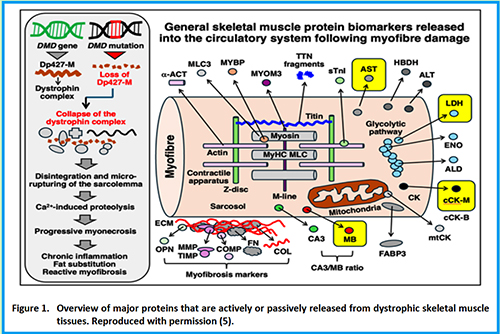
5. Dowling P, Negroni E, Trollet C, Zweyer M, Swandulla D, Ohlendieck K. Serum protein biomarker signature of Duchenne muscular dystrophy. Eur J Transl Myol. 2025 Jun 27;35(2):13956. doi: 10.4081/ejtm.2025.13956. Epub 2025 May 28. PMID: 40438995; PMCID: PMC12265423.

How to Cite


1.
Ohlendieck K. Abstract 025 | Proteomic profiling of the biofluid marker signature of dystrophinopathies: Kay Ohlendieck 1|2, Dieter Swandulla 3, Paul Dowling 1|2 | 1Department of Biology, Maynooth University, Maynooth, Co. Kildare, Ireland; 2Kathleen Lonsdale Institute for Human Health Research, Maynooth University, Maynooth, Co. Kildare, Ireland; 3Institute of Physiology, Faculty of Medicine, University of Bonn, Bonn, Germany. Eur J Transl Myol [Internet]. 2026 Mar. 2 [cited 2026 Apr. 17];36(s1). Available from: https://www.pagepressjournals.org/bam/article/view/15024