Validity of ultrasound rectus femoris quantitative assessment: A comparative study between linear and curved array transducers
All claims expressed in this article are solely those of the authors and do not necessarily represent those of their affiliated organizations, or those of the publisher, the editors and the reviewers. Any product that may be evaluated in this article or claim that may be made by its manufacturer is not guaranteed or endorsed by the publisher.
Published: 7 December 2022
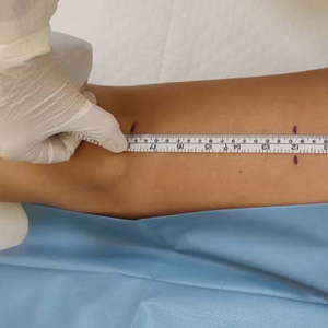
Appendicular skeletal mass is commonly used to assess the loss in muscle mass and US represents a valid, and reliable method. However, the procedural protocols are still heterogeneous. The aim of this study was to compare the intertransducers validity of thickness, width, and CSA measurements of RF muscle. The AP, LL and CSA of RF muscle were evaluated with both linear and curve probes in ten healthy subjects and six sarcopenic patients. In the healthy group the mean AP diameters measured with the linear array were significantly higher than those measured with the curved array. AP and CSA were higher in the healthy group compared with the sarcopenic group with both transducers. There was a positive correlation between weight and LL diameter, and a negative correlation between age and muscle AP, measured with the linear probe. Both linear and curved probes represent valid methods in US evaluation of the CSA of the RF muscle. However, in the healthy subjects, the thickness and width of the of the same muscle, are affected by the type of probe.
Downloads
Anton SD, Woods AJ, Ashizawa T, Barb D, Buford TW, Carter CS, Clark DJ, Cohen RA, Corbett DB, Cruz-Almeida Y, Dotson V, Ebner N, Efron PA, Fillingim RB, Foster TC, Gundermann DM, Joseph AM, Karabetian C, Leeuwenburgh C, Manini TM, Marsiske M, Mankowski RT, Mutchie HL, Perri MG, Ranka S, Rashidi P, Sandesara B, Scarpace PJ, Sibille KT, Solberg LM, Someya S, Uphold C, Wohlgemuth S, Wu SS, Pahor M. Successful aging: Advancing the science of physical independence in older adults. Ageing Res Rev. 2015 Nov;24(Pt B):304-27. Epub 2015 Oct 14. DOI: https://doi.org/10.1016/j.arr.2015.09.005
Marcell TJ. Sarcopenia: causes, consequences, and preventions. J Gerontol A Biol Sci Med Sci. 2003 Oct;58(10):M911-6. DOI: https://doi.org/10.1093/gerona/58.10.M911
Coletti D, Daou N, Hassani M, Li Z, Parlakian A. Serum Response Factor in Muscle Tissues: From Development to Ageing. Eur J Transl Myol. 2016 Jun 22;26(2):6008. DOI: https://doi.org/10.4081/ejtm.2016.6008
Wang JC, Wu WT, Chang KV, Chen LR, Chi SY, Kara M ÖL. Ultrasound Imaging for the Diagnosis and Evaluation of Sarcopenia: An Umbrella Review. Life. 2021;12(1):9. DOI: https://doi.org/10.3390/life12010009
Hellmanns, K.; McBean K. Magnetic Resonance Imaging in the measurement of whole body muscle mass: A comparison of interval gap methods. Radiography. 2015;21:e35–e39. DOI: https://doi.org/10.1016/j.radi.2014.09.009
Cao, Q.; Xiong, Y.; Zhong, Z.; Ye Q. Computed Tomography-Assessed Sarcopenia Indexes Predict Major Complications following Surgery for Hepatopancreatobiliary Malignancy: A Meta-Analysis. Ann Nutr Metab. 2019;74:24–34. DOI: https://doi.org/10.1159/000494887
Buckinx, F.; Landi, F.; Cesari, M.; Fielding, R.A.; Visser, M.; Engelke, K.; Maggi, S.; Dennison, E.; Al-Daghri, N.M.; Allepaerts S., Al E. Pitfalls in the measurement of muscle mass: A need for a reference standard. J Cachexia Sarcopenia Muscle. 2018;9:269–278. DOI: https://doi.org/10.1002/jcsm.12268
Lam, F.M.H.; Su, Y.; Lu, Z.H.; Yu, R.; Leung, J.C.S.; Kwok TC. Cumulative and Incremental Value of Sarcopenia Components on Predicting Adverse Outcomes. J Am Med Dir Assoc. 2020;21:1481–9. DOI: https://doi.org/10.1016/j.jamda.2020.05.056
Larsson L, Degens H, Li M, Salviati L, Lee YI, Thompson W, Kirkland JL, Sandri M. Sarcopenia: Aging-related loss of muscle mass and function. Physiol Rev. 2019;99(1):427–511. DOI: https://doi.org/10.1152/physrev.00061.2017
Kara M, Kaymak B, Frontera W, Ata AM, Ricci V, Ekiz T, Chang KV, Han DS, Michail X, Quittan M, Lim JY, Bean JF, Franchignoni F, Özçakar L. Diagnosing sarcopenia: Functional perspectives and a new algorithm from ISarcoPRM. J Rehabil Med. 2021;53(6). DOI: https://doi.org/10.2340/16501977-2851
Abe T, Patterson KM, Stover CD, Geddam DA, Tribby AC, Lajza DG, Young KC. Site-specific thigh muscle loss as an independent phenomenon for age-related muscle loss in middle-aged and older men and women. Age (Omaha). 2014;36(3):1353–8. DOI: https://doi.org/10.1007/s11357-014-9634-3
Dehghan M, Merchant A. Is bioelectrical impedance accurate for use in large epidemiological studies? Nutr J. 2008;7(26). DOI: https://doi.org/10.1186/1475-2891-7-26
Berger J, Bunout D, Barrera G, de la Maza MP, Henriquez S, Leiva L, Hirsch S. Rectus femoris (RF) ultrasound for the assessment of muscle mass in older people. Arch Gerontol Geriatr. 2015;61(1):33–8. DOI: https://doi.org/10.1016/j.archger.2015.03.006
Nijholt W, Scafoglieri A, Jager-Wittenaar H, Hobbelen JSM, van der Schans CP. The reliability and validity of ultrasound to quantify muscles in older adults: a systematic review. J Cachexia Sarcopenia Muscle. 2017;8(5):702–12. DOI: https://doi.org/10.1002/jcsm.12210
Hammond K, Mampilly J, Laghi FA, Goyal A, Collins EG, McBurney C, Jubran A, Tobin MJ. Validity and reliability of rectus femoris ultrasound measurements: Comparison of curved-array and linear-array transducers. J Rehabil Res Dev. 2014;51(7):1155–64. DOI: https://doi.org/10.1682/JRRD.2013.08.0187
Ticinesi A, Meschi T, Narici M V., Lauretani F, Maggio M. Muscle Ultrasound and Sarcopenia in Older Individuals: A Clinical Perspective. J Am Med Dir Assoc [Internet]. 2017;18(4):290–300. DOI: https://doi.org/10.1016/j.jamda.2016.11.013
Strasser EM, Draskovits T, Praschak M, Quittan M, Graf A. Association between ultrasound measurements of muscle thickness, pennation angle, echogenicity and skeletal muscle strength in the elderly. Age (Omaha). 2013;35(6):2377–88. DOI: https://doi.org/10.1007/s11357-013-9517-z
Cruz-Jentoft AJ, Bahat G, Bauer J, Boirie Y, Bruyère O, Cederholm T, Cooper C, Landi F, Rolland Y, Sayer AA, Schneider SM, Sieber CC, Topinkova E, Vandewoude M, Visser M, Zamboni M. Sarcopenia: Revised European consensus on definition and diagnosis. Age Ageing. 2019;48(1):16–31. DOI: https://doi.org/10.1093/ageing/afy169
Coraci D, Giovannini S, Loreti C, Ruggeri F PL. The hyperchoic rim of the normal nerve in ultrasound: how significant is it? Neurol Sci. 2020;41(10):2985–7. DOI: https://doi.org/10.1007/s10072-020-04405-6
Adamec C. Pouzití Spearmanova koeficientu korelace poradí pri validaci [The use of Spearman's correlation order coefficient in validation]. Cesk Zdrav. 1966 Feb;14(2):100-2. Czech.
Ozcakar L, De Muynck Martine. Musculoskeletal Ultrasound in Physical and Rehabilitation Medicine. 1st Ed. Edi Ermes. 2014. p. 11–15.
Gonzalez A, Valero-Breton M, Huerta-Salgado C, Achiardi O, Simon F C-VC. Impact of exercise training on the sarcopenia criteria in non-alcoholic fatty liver disease: a systematic review and meta-analysis. Eur J Transl Myol. 2021;31(1):9630. DOI: https://doi.org/10.4081/ejtm.2020.9630
Perkisas S, Bastijns S, Sanchez-Rodriguez D, Piotrowicz K, De Cock AM; full SARCUS working group.Application of ultrasound for muscle assessment in sarcopenia: 2020 SARCUS update. Eur Geriatr Med [Internet]. 2021;12(1):45–59. DOI: https://doi.org/10.1007/s41999-020-00433-9
Baumgartner RN. Body composition in healthy aging. Ann N Y Acad Sci. 2000;904:437–48. DOI: https://doi.org/10.1111/j.1749-6632.2000.tb06498.x
Kim S, Won CW. Sex-different changes of body composition in aging: a systemic review. Arch Gerontol Geriatr [Internet]. 2022;102 (May): 104711. DOI: https://doi.org/10.1016/j.archger.2022.104711
Mitchell W, Williams J, Atherton P, Larvin M, Lund J, Narici M. Sarcopenia, dynapenia, and the impact of advancing age on human skeletal muscle size and strength; a quantitative review. Front Physiol. 2012;(July):1–18. DOI: https://doi.org/10.3389/fphys.2012.00260
Blazevich AJ, Cannavan D, Coleman DR, Horne S. Influence of concentric and eccentric resistance training on architectural adaptation in human quadriceps muscles. J Appl Physiol. 2007;103(5):1565–75. DOI: https://doi.org/10.1152/japplphysiol.00578.2007
Alegre LM, Jiménez F, Gonzalo-Orden JM, Martín-Acero R AX. Effects of dynamic resistance training on fascicle length and isometric strength. J Sport Sci. 2006;24(5):501–8. DOI: https://doi.org/10.1080/02640410500189322
Reeves ND, Narici MV MC. In vivo human muscle structure and function: adaptations to resistance training in old age. Exp Physiol. 2004;89(6):675–89. DOI: https://doi.org/10.1113/expphysiol.2004.027797
Seynnes OR, de Boer M NM. Early skeletal muscle hypertrophy and architectural changes in response to high-intensity resistance training. J Appl Physiol. 2007;102(1):368–73. DOI: https://doi.org/10.1152/japplphysiol.00789.2006
Cruz-Jentoft AJ, Bahat G, Bauer J, Boirie Y, Bruyère O, Cederholm T, Cooper C, Landi F, Rolland Y, Sayer AA, Schneider SM, Sieber CC, Topinkova E, Vandewoude M, Visser M, Zamboni M; Writing Group for the European Working Group on Sarcopenia in Older People 2 (EWGSOP2), and the Extended Group for EWGSOP2. Sarcopenia: Revised European consensus on definition and diagnosis. Eur Geriatr Med [Internet]. 2021;12(1):1–13.
Aagaard P, Suetta C, Caserotti P, Magnusson SP, Kjær M. Role of the nervous system in sarcopenia and muscle atrophy with aging: Strength training as a countermeasure. Scand J Med Sci Sport. 2010;20(1):49–64. DOI: https://doi.org/10.1111/j.1600-0838.2009.01084.x
Baumgartner RN, Koehler KM, Gallagher D, Romero L, Heymsfield SB, Ross RR, Garry PJ LR. Epidemiology of sarcopenia among the elderly in New Mexico. Am J Epidemiol. 1998; 147 (8): 755–63. DOI: https://doi.org/10.1093/oxfordjournals.aje.a009520
Burns JM, Johnson DK, Watts A, Swerdlow RH BW. Reduced lean mass in early Alzheimer disease and its association with brain atrophy. Arch Neurol. 2010;67(4):428–33. DOI: https://doi.org/10.1001/archneurol.2010.38
Park SW, Goodpaster BH, Lee JS, Kuller LH, Boudreau R, de Rekeneire N, Harris TB, Kritchevsky S, Tylavsky FA, Nevitt M, Cho YW NA. Health, Aging, and Body Composition Study. Excessive loss of skeletal muscle mass in older adults with type 2 diabetes. Diabetes Care. 2009;32(11):1993–7. DOI: https://doi.org/10.2337/dc09-0264
How to Cite

This work is licensed under a Creative Commons Attribution-NonCommercial 4.0 International License.
PAGEPress has chosen to apply the Creative Commons Attribution NonCommercial 4.0 International License (CC BY-NC 4.0) to all manuscripts to be published.