
Termination of the great saphenous vein at variable levels
All claims expressed in this article are solely those of the authors and do not necessarily represent those of their affiliated organizations, or those of the publisher, the editors and the reviewers. Any product that may be evaluated in this article or claim that may be made by its manufacturer is not guaranteed or endorsed by the publisher.
Published: 22 December 2022
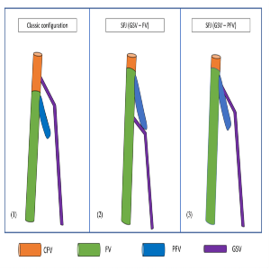
The assessment of the saphenofemoral junction (SFJ) is important in the diagnosis and treatment of venous reflux of the great saphenous vein (GSV). In the clinical practice of venous medicine, the SFJ is used to represent the region at which the saphenous arch connects with the common femoral vein (CFV). A number of notable variations of the SFJ have been documented, and rare variable courses of the GSV have been described recently. Our case study reports two unusual GSV terminations. In both cases, the SFJ was located below the confluence of the profunda femoris vein (PFV) with the femoral vein (FV). Case 1 showed the SFJ was formed by the GSV and FV; whereas case 2 showed the PFV was joined by the GSV after a transposition with the FV. Anatomical variations of the SFJ are rare; however, they are increasingly diagnosed with the use of duplex ultrasound. The identification of SFJ variants warrants a safe endovenous procedure and prevents surgical complications.
Downloads
Caggiati A, Bergan JJ, Gloviczki P, et al. Nomenclature of the veins of the lower limb: extensions, refinements, and clinical application. J Vasc Surg. 2005;41:719-24. DOI: https://doi.org/10.1016/j.jvs.2005.01.018
Souroullas P, Barnes R, Smith G, et al. The classic saphenofemoral junction and its anatomical variations. Phlebology. 2017;32:172-8. DOI: https://doi.org/10.1177/0268355516635960
Igari K, Hirokawa M, Uchiyama H, et al. Anatomical variation at the sapheno-femoral junction. Ann Vasc Dis. 2013;6:702-5. DOI: https://doi.org/10.3400/avd.oa.13-00087
Quickert T, Alagha M. A rare anatomical variation of great saphenous vein at the level of saphenofemoral junction. Radiol Case Rep. 2018;13:1128-9. DOI: https://doi.org/10.1016/j.radcr.2018.07.028
Lekich C, Campbell W, Walton S, Hannah P. Anomalous high bifurcation of the common femoral artery at the groin: management of GSV incompetence with endovenous laser ablation. Phlebology. 2013;28:264-7. DOI: https://doi.org/10.1258/phleb.2012.012047
Nabatoff RA. Anomalies encountered during varicose vein surgery. Arch Surg. 1978;113:586-8. DOI: https://doi.org/10.1001/archsurg.1978.01370170048007
Uhl JF, Gillot C. Anatomy and embryology of the small saphenous vein: nerve relationships and implications for treatment. Phlebology. 2013;28:4-15. DOI: https://doi.org/10.1258/phleb.2012.012j08
Coleridge-Smith P, Labropoulos N, Partsch H, et al. Duplex ultrasound investigation of the veins in chronic venous disease of the lower limbs-UIP consensus document. Part I. Basic principles. Eur J Vasc Endovasc Surg. 2006;31:83-92. DOI: https://doi.org/10.1016/j.ejvs.2005.07.019
Kabnick LS, Sadek M, Bjarnason H, et al. Classification and treatment of endothermal heat-induced thrombosis: Recommendations from the American Venous Forum and the Society for Vascular Surgery. Phlebology. 2021;36:8-25. DOI: https://doi.org/10.1177/0268355520953759
Kim DS, Kim SW, Lee HS, et al. Rare Vascular Anomalies in the Femoral Triangle During Varicose Vein Surgery. Korean J Thorac Cardiovasc Surg. 2017;50:99-104. DOI: https://doi.org/10.5090/kjtcs.2017.50.2.99
Supporting Agencies
How to Cite

This work is licensed under a Creative Commons Attribution-NonCommercial 4.0 International License.
PAGEPress has chosen to apply the Creative Commons Attribution NonCommercial 4.0 International License (CC BY-NC 4.0) to all manuscripts to be published.

