Involvement of muscle satellite cell dysfunction in neuromuscular disorders: Expanding the portfolio of satellite cell-opathies
All claims expressed in this article are solely those of the authors and do not necessarily represent those of their affiliated organizations, or those of the publisher, the editors and the reviewers. Any product that may be evaluated in this article or claim that may be made by its manufacturer is not guaranteed or endorsed by the publisher.
Published: 18 March 2022
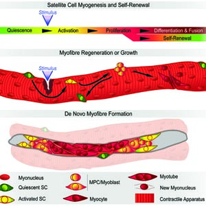
Neuromuscular disorders are a heterogeneous group of acquired or hereditary conditions that affect striated muscle function. The resulting decrease in muscle strength and motility irreversibly impacts quality of life. In addition to directly affecting skeletal muscle, pathogenesis can also arise from dysfunctional crosstalk between nerves and muscles, and may include cardiac impairment. Muscular weakness is often progressive and paralleled by continuous decline in the ability of skeletal muscle to functionally adapt and regenerate. Normally, the skeletal muscle resident stem cells, named satellite cells, ensure tissue homeostasis by providing myoblasts for growth, maintenance, repair and regeneration. We recently defined 'Satellite Cell-opathies’ as those inherited neuromuscular conditions presenting satellite cell dysfunction in muscular dystrophies and myopathies (doi:10.1016/j.yexcr.2021.112906). Here, we expand the portfolio of Satellite Cell-opathies by evaluating the potential impairment of satellite cell function across all 16 categories of neuromuscular disorders, including those with mainly neurogenic and cardiac involvement. We explore the expression dynamics of myopathogenes, genes whose mutation leads to skeletal muscle pathogenesis, using transcriptomic analysis. This revealed that 45% of myopathogenes are differentially expressed during early satellite cell activation (0 - 5 hours). Of these 271 myopathogenes, 83 respond to Pax7, a master regulator of satellite cells. Our analysis suggests possible perturbation of satellite cell function in many neuromuscular disorders across all categories, including those where skeletal muscle pathology is not predominant. This characterisation further aids understanding of pathomechanisms and informs on development of prognostic and diagnostic tools, and ultimately, new therapeutics.
Downloads
Mukund K, Subramaniam S. Skeletal muscle: A review of molecular structure and function, in health and disease. Wiley Interdiscip Rev Syst Biol Med. 2020;12(1):e1462. Epub 2019/08/14. DOI: https://doi.org/10.1002/wsbm.1462
Dowling JJ, Weihl CC, Spencer MJ. Molecular and cellular basis of genetically inherited skeletal muscle disorders. Nat Rev Mol Cell Biol. 2021. Epub 2021/07/15.
Ganassi M, Muntoni F, Zammit PS. Defining and identifying satellite cell-opathies within muscular dystrophies and myopathies. Exp Cell Res. 2022;411(1):112906. Epub 2021/11/07. DOI: https://doi.org/10.1016/j.yexcr.2021.112906
Benarroch L, Bonne G, Rivier F, Hamroun D. The 2021 version of the gene table of neuromuscular disorders (nuclear genome). Neuromuscul Disord. 2020;30(12):1008-48. Epub 2020/12/02. DOI: https://doi.org/10.1016/j.nmd.2020.11.009
Fukada SI, Akimoto T, Sotiropoulos A. Role of damage and management in muscle hypertrophy: Different behaviors of muscle stem cells in regeneration and hypertrophy. Biochim Biophys Acta Mol Cell Res. 2020;1867(9):118742. Epub 2020/05/18. DOI: https://doi.org/10.1016/j.bbamcr.2020.118742
Mauro A. Satellite cell of skeletal muscle fibers. J Biophys Biochem Cytol. 1961;9:493-5. Epub 1961/02/01. DOI: https://doi.org/10.1083/jcb.9.2.493
Relaix F, Zammit PS. Satellite cells are essential for skeletal muscle regeneration: the cell on the edge returns centre stage. Development. 2012;139(16):2845-56. Epub 2012/07/27. DOI: https://doi.org/10.1242/dev.069088
Cardamone M, Darras BT, Ryan MM. Inherited myopathies and muscular dystrophies. Semin Neurol. 2008;28(2):250-9. Epub 2008/03/21. DOI: https://doi.org/10.1055/s-2008-1062269
Sewry CA, Wallgren-Pettersson C. Myopathology in congenital myopathies. Neuropathol Appl Neurobiol. 2017;43(1):5-23. Epub 2016/12/16. DOI: https://doi.org/10.1111/nan.12369
Collins CA, Olsen I, Zammit PS, Heslop L, Petrie A, Partridge TA, et al. Stem cell function, self-renewal, and behavioral heterogeneity of cells from the adult muscle satellite cell niche. Cell. 2005;122(2):289-301. Epub 2005/07/30. DOI: https://doi.org/10.1016/j.cell.2005.05.010
Collins CA, Partridge TA. Self-renewal of the adult skeletal muscle satellite cell. Cell Cycle. 2005;4(10):1338-41. Epub 2005/09/24. DOI: https://doi.org/10.4161/cc.4.10.2114
Moss FP, Leblond CP. Satellite cells as the source of nuclei in muscles of growing rats. Anat Rec. 1971;170(4):421-35. Epub 1971/08/01. DOI: https://doi.org/10.1002/ar.1091700405
Morgan J, Butler-Browne G, Muntoni F, Patel K, skeletal muscle stem cells involvement in pathology study g. 240th ENMC workshop: The involvement of skeletal muscle stem cells in the pathology of muscular dystrophies 25-27 January 2019, Hoofddorp, The Netherlands. Neuromuscul Disord. 2019;29(9):704-15. Epub 2019/08/27. DOI: https://doi.org/10.1016/j.nmd.2019.07.003
Lopes F, Miguet M, Mucha BE, Gauthier J, Saillour V, Nguyen CE, et al. MYOD1 involvement in myopathy. Eur J Neurol. 2018;25(12):e123-e4. Epub 2018/11/08. DOI: https://doi.org/10.1111/ene.13782
Shukla A, Narayanan DL, Asher U, Girisha KM. A novel bi-allelic loss-of-function variant in MYOD1: Further evidence for gene-disease association and phenotypic variability in MYOD1-related myopathy. Clin Genet. 2019;96(3):276-7. Epub 2019/07/02. DOI: https://doi.org/10.1111/cge.13596
Watson CM, Crinnion LA, Murphy H, Newbould M, Harrison SM, Lascelles C, et al. Deficiency of the myogenic factor MyoD causes a perinatally lethal fetal akinesia. J Med Genet. 2016;53(4):264-9. Epub 2016/01/07. doi: 10.1136/jmedgenet-2015-103620. DOI: https://doi.org/10.1136/jmedgenet-2015-103620
Banerji CRS, Henderson D, Tawil RN, Zammit PS. Skeletal muscle regeneration in facioscapulohumeral muscular dystrophy is correlated with pathological severity. Hum Mol Genet. 2020;29(16):2746-60. Epub 2020/08/04. DOI: https://doi.org/10.1093/hmg/ddaa164
Hewitt JE, Lyle R, Clark LN, Valleley EM, Wright TJ, Wijmenga C, et al. Analysis of the tandem repeat locus D4Z4 associated with facioscapulohumeral muscular dystrophy. Hum Mol Genet. 1994;3(8):1287-95. Epub 1994/08/01. DOI: https://doi.org/10.1093/hmg/3.8.1287
van Deutekom JC, Wijmenga C, van Tienhoven EA, Gruter AM, Hewitt JE, Padberg GW, et al. FSHD associated DNA rearrangements are due to deletions of integral copies of a 3.2 kb tandemly repeated unit. Hum Mol Genet. 1993;2(12):2037-42. Epub 1993/12/01. DOI: https://doi.org/10.1093/hmg/2.12.2037
Greco A, Goossens R, van Engelen B, van der Maarel SM. Consequences of epigenetic derepression in facioscapulohumeral muscular dystrophy. Clin Genet. 2020;97(6):799-814. Epub 2020/02/23. DOI: https://doi.org/10.1111/cge.13726
Lim KRQ, Nguyen Q, Yokota T. DUX4 Signalling in the Pathogenesis of Facioscapulohumeral Muscular Dystrophy. Int J Mol Sci. 2020;21(3). Epub 2020/01/26. DOI: https://doi.org/10.3390/ijms21030729
Heher P, Ganassi M, Weidinger A, Engquist EN, Pruller J, Nguyen TH, et al. Interplay between mitochondrial reactive oxygen species, oxidative stress and hypoxic adaptation in facioscapulohumeral muscular dystrophy: Metabolic stress as potential therapeutic target. Redox Biology. 2022:102251. DOI: https://doi.org/10.1016/j.redox.2022.102251
Banerji CRS, Panamarova M, Hebaishi H, White RB, Relaix F, Severini S, et al. PAX7 target genes are globally repressed in facioscapulohumeral muscular dystrophy skeletal muscle. Nat Commun. 2017;8(1):2152. Epub 2017/12/20. DOI: https://doi.org/10.1038/s41467-017-01200-4
Banerji CRS, Zammit PS. PAX7 target gene repression is a superior FSHD biomarker than DUX4 target gene activation, associating with pathological severity and identifying FSHD at the single-cell level. Hum Mol Genet. 2019;28(13):2224-36. Epub 2019/05/09. DOI: https://doi.org/10.1093/hmg/ddz043
Bosnakovski D, Toso EA, Hartweck LM, Magli A, Lee HA, Thompson ER, et al. The DUX4 homeodomains mediate inhibition of myogenesis and are functionally exchangeable with the Pax7 homeodomain. J Cell Sci. 2017;130(21):3685-97. Epub 2017/09/25. DOI: https://doi.org/10.1242/jcs.205427
Omar A, Marwaha K, Bollu PC. Physiology, Neuromuscular Junction. StatPearls. Treasure Island (FL)2021.
Jimsheleishvili S, Marwaha K, Sherman A. Physiology, Neuromuscular Transmission. StatPearls. Treasure Island (FL)2021.
Kelly AM. Perisynaptic satellite cells in the developing and mature rat soleus muscle. Anat Rec. 1978;190(4):891-903. Epub 1978/04/01. DOI: https://doi.org/10.1002/ar.1091900409
Liu W, Wei-LaPierre L, Klose A, Dirksen RT, Chakkalakal JV. Inducible depletion of adult skeletal muscle stem cells impairs the regeneration of neuromuscular junctions. Elife. 2015;4. Epub 2015/08/28. DOI: https://doi.org/10.7554/eLife.09221
Sanes JR, Marshall LM, McMahan UJ. Reinnervation of muscle fiber basal lamina after removal of myofibers. Differentiation of regenerating axons at original synaptic sites. J Cell Biol. 1978;78(1):176-98. Epub 1978/07/01. DOI: https://doi.org/10.1083/jcb.78.1.176
Bruusgaard JC, Gundersen K. In vivo time-lapse microscopy reveals no loss of murine myonuclei during weeks of muscle atrophy. J Clin Invest. 2008;118(4):1450-7. Epub 2008/03/05. DOI: https://doi.org/10.1172/JCI34022
Scaramozza A, Marchese V, Papa V, Salaroli R, Soraru G, Angelini C, et al. Skeletal muscle satellite cells in amyotrophic lateral sclerosis. Ultrastruct Pathol. 2014;38(5):295-302. Epub 2014/08/01. DOI: https://doi.org/10.3109/01913123.2014.937842
Burr P, Reddivari AKR. Spinal Muscle Atrophy. StatPearls. Treasure Island (FL)2021.
McCormack NM, Villalon E, Viollet C, Soltis AR, Dalgard CL, Lorson CL, et al. Survival motor neuron deficiency slows myoblast fusion through reduced myomaker and myomixer expression. J Cachexia Sarcopenia Muscle. 2021;12(4):1098-116. Epub 2021/06/12. DOI: https://doi.org/10.1002/jcsm.12740
Hellbach N, Peterson S, Haehnke D, Shankar A, LaBarge S, Pivaroff C, et al. Impaired myogenic development, differentiation and function in hESC-derived SMA myoblasts and myotubes. PLoS One. 2018;13(10):e0205589. Epub 2018/10/12. DOI: https://doi.org/10.1371/journal.pone.0205589
Bushby K, Muntoni F, Bourke JP. 107th ENMC international workshop: the management of cardiac involvement in muscular dystrophy and myotonic dystrophy. 7th-9th June 2002, Naarden, the Netherlands. Neuromuscul Disord. 2003;13(2):166-72. Epub 2003/02/05. DOI: https://doi.org/10.1016/S0960-8966(02)00213-4
Muntoni F. Cardiac complications of childhood myopathies. J Child Neurol. 2003;18(3):191-202. Epub 2003/05/07. DOI: https://doi.org/10.1177/08830738030180030301
Verhaert D, Richards K, Rafael-Fortney JA, Raman SV. Cardiac involvement in patients with muscular dystrophies: magnetic resonance imaging phenotype and genotypic considerations. Circ Cardiovasc Imaging. 2011;4(1):67-76. Epub 2011/01/20. DOI: https://doi.org/10.1161/CIRCIMAGING.110.960740
Kostareva A, Sejersen T, Sjoberg G. Genetic spectrum of cardiomyopathies with neuromuscular phenotype. Front Biosci (Schol Ed). 2013;5:325-40. Epub 2013/01/02. DOI: https://doi.org/10.2741/S375
Ripa R, George T, Sattar Y. Physiology, Cardiac Muscle. StatPearls. Treasure Island (FL)2021.
Sweeney HL, Hammers DW. Muscle Contraction. Cold Spring Harb Perspect Biol. 2018;10(2). Epub 2018/02/09. DOI: https://doi.org/10.1101/cshperspect.a023200
Machado L, Esteves de Lima J, Fabre O, Proux C, Legendre R, Szegedi A, et al. In Situ Fixation Redefines Quiescence and Early Activation of Skeletal Muscle Stem Cells. Cell Rep. 2017;21(7):1982-93. Epub 2017/11/16. DOI: https://doi.org/10.1016/j.celrep.2017.10.080
Relaix F, Rocancourt D, Mansouri A, Buckingham M. Divergent functions of murine Pax3 and Pax7 in limb muscle development. Genes Dev. 2004;18(9):1088-105. Epub 2004/05/11. DOI: https://doi.org/10.1101/gad.301004
Seale P, Sabourin LA, Girgis-Gabardo A, Mansouri A, Gruss P, Rudnicki MA. Pax7 is required for the specification of myogenic satellite cells. Cell. 2000;102(6):777-86. Epub 2000/10/13. DOI: https://doi.org/10.1016/S0092-8674(00)00066-0
Zammit PS, Relaix F, Nagata Y, Ruiz AP, Collins CA, Partridge TA, et al. Pax7 and myogenic progression in skeletal muscle satellite cells. J Cell Sci. 2006;119(Pt 9):1824-32. Epub 2006/04/13. DOI: https://doi.org/10.1242/jcs.02908
Lilja KC, Zhang N, Magli A, Gunduz V, Bowman CJ, Arpke RW, et al. Pax7 remodels the chromatin landscape in skeletal muscle stem cells. PLoS One. 2017;12(4):e0176190. Epub 2017/04/26. DOI: https://doi.org/10.1371/journal.pone.0176190
Jain A, Al Khalili Y. Congenital Myotonic Dystrophy. StatPearls. Treasure Island (FL)2022.
Thornell LE, Lindstom M, Renault V, Klein A, Mouly V, Ansved T, et al. Satellite cell dysfunction contributes to the progressive muscle atrophy in myotonic dystrophy type 1. Neuropathol Appl Neurobiol. 2009;35(6):603-13. Epub 2009/02/12. DOI: https://doi.org/10.1111/j.1365-2990.2009.01014.x
Furling D, Coiffier L, Mouly V, Barbet JP, St Guily JL, Taneja K, et al. Defective satellite cells in congenital myotonic dystrophy. Hum Mol Genet. 2001;10(19):2079-87. Epub 2001/10/09. DOI: https://doi.org/10.1093/hmg/10.19.2079
Iyer SR, Shah SB, Lovering RM. The Neuromuscular Junction: Roles in Aging and Neuromuscular Disease. Int J Mol Sci. 2021;22(15). Epub 2021/08/08. DOI: https://doi.org/10.3390/ijms22158058
Ng SY, Ljubicic V. Recent insights into neuromuscular junction biology in Duchenne muscular dystrophy: Impacts, challenges, and opportunities. EBioMedicine. 2020;61:103032. Epub 2020/10/12. DOI: https://doi.org/10.1016/j.ebiom.2020.103032
Michele DE, Barresi R, Kanagawa M, Saito F, Cohn RD, Satz JS, et al. Post-translational disruption of dystroglycan-ligand interactions in congenital muscular dystrophies. Nature. 2002;418(6896):417-22. Epub 2002/07/26. DOI: https://doi.org/10.1038/nature00837
Brancaccio A. A molecular overview of the primary dystroglycanopathies. J Cell Mol Med. 2019;23(5):3058-62. Epub 2019/03/07. DOI: https://doi.org/10.1111/jcmm.14218
Gonzalez-Perez P, Smith C, Sebetka WL, Gedlinske A, Perlman S, Mathews KD. Clinical and electrophysiological evaluation of myasthenic features in an alpha-dystroglycanopathy cohort (FKRP-predominant). Neuromuscul Disord. 2020;30(3):213-8. Epub 2020/03/03. DOI: https://doi.org/10.1016/j.nmd.2020.01.002
Cohn RD, Henry MD, Michele DE, Barresi R, Saito F, Moore SA, et al. Disruption of DAG1 in differentiated skeletal muscle reveals a role for dystroglycan in muscle regeneration. Cell. 2002;110(5):639-48. Epub 2002/09/17. DOI: https://doi.org/10.1016/S0092-8674(02)00907-8
Beedle AM, Turner AJ, Saito Y, Lueck JD, Foltz SJ, Fortunato MJ, et al. Mouse fukutin deletion impairs dystroglycan processing and recapitulates muscular dystrophy. J Clin Invest. 2012;122(9):3330-42. Epub 2012/08/28. DOI: https://doi.org/10.1172/JCI63004
Saito F, Masaki T, Saito Y, Nakamura A, Takeda S, Shimizu T, et al. Defective peripheral nerve myelination and neuromuscular junction formation in fukutin-deficient chimeric mice. J Neurochem. 2007;101(6):1712-22. Epub 2007/03/01. DOI: https://doi.org/10.1111/j.1471-4159.2007.04462.x
Hiroi A, Yamamoto T, Shibata N, Osawa M, Kobayashi M. Roles of fukutin, the gene responsible for fukuyama-type congenital muscular dystrophy, in neurons: possible involvement in synaptic function and neuronal migration. Acta Histochem Cytochem. 2011;44(2):91-101. Epub 2011/05/27. DOI: https://doi.org/10.1267/ahc.10045
Taniguchi M, Kurahashi H, Noguchi S, Fukudome T, Okinaga T, Tsukahara T, et al. Aberrant neuromuscular junctions and delayed terminal muscle fiber maturation in alpha-dystroglycanopathies. Hum Mol Genet. 2006;15(8):1279-89. Epub 2006/03/15. DOI: https://doi.org/10.1093/hmg/ddl045
Arimura T, Hayashi YK, Murakami T, Oya Y, Funabe S, Arikawa-Hirasawa E, et al. Mutational analysis of fukutin gene in dilated cardiomyopathy and hypertrophic cardiomyopathy. Circ J. 2009;73(1):158-61. Epub 2008/11/19. DOI: https://doi.org/10.1253/circj.CJ-08-0722
Servian-Morilla E, Cabrera-Serrano M, Johnson K, Pandey A, Ito A, Rivas E, et al. POGLUT1 biallelic mutations cause myopathy with reduced satellite cells, alpha-dystroglycan hypoglycosylation and a distinctive radiological pattern. Acta Neuropathol. 2020;139(3):565-82. Epub 2020/01/04. DOI: https://doi.org/10.1007/s00401-019-02117-6
Servian-Morilla E, Takeuchi H, Lee TV, Clarimon J, Mavillard F, Area-Gomez E, et al. A POGLUT1 mutation causes a muscular dystrophy with reduced Notch signaling and satellite cell loss. EMBO Mol Med. 2016;8(11):1289-309. Epub 2016/11/04. DOI: https://doi.org/10.15252/emmm.201505815
Ribeiro AF, Jr., Souza LS, Almeida CF, Ishiba R, Fernandes SA, Guerrieri DA, et al. Muscle satellite cells and impaired late stage regeneration in different murine models for muscular dystrophies. Sci Rep. 2019;9(1):11842. Epub 2019/08/16. DOI: https://doi.org/10.1038/s41598-019-48156-7
Hoffman EP, Brown RH, Jr., Kunkel LM. Dystrophin: the protein product of the Duchenne muscular dystrophy locus. Cell. 1987;51(6):919-28. Epub 1987/12/24. DOI: https://doi.org/10.1016/0092-8674(87)90579-4
Zhang M, McLennan IS. Use of antibodies to identify satellite cells with a light microscope. Muscle Nerve. 1994;17(9):987-94. Epub 1994/09/01. DOI: https://doi.org/10.1002/mus.880170905
Dumont NA, Wang YX, von Maltzahn J, Pasut A, Bentzinger CF, Brun CE, et al. Dystrophin expression in muscle stem cells regulates their polarity and asymmetric division. Nat Med. 2015;21(12):1455-63. Epub 2015/11/17. DOI: https://doi.org/10.1038/nm.3990
Kong J, Anderson JE. Dystrophin is required for organizing large acetylcholine receptor aggregates. Brain Res. 1999;839(2):298-304. Epub 1999/10/16. DOI: https://doi.org/10.1016/S0006-8993(99)01737-0
Sancar F, Touroutine D, Gao S, Oh HJ, Gendrel M, Bessereau JL, et al. The dystrophin-associated protein complex maintains muscle excitability by regulating Ca(2+)-dependent K(+) (BK) channel localization. J Biol Chem. 2011;286(38):33501-10. Epub 2011/07/29. DOI: https://doi.org/10.1074/jbc.M111.227678
Kottlors M, Kirschner J. Elevated satellite cell number in Duchenne muscular dystrophy. Cell Tissue Res. 2010;340(3):541-8. Epub 2010/05/15. DOI: https://doi.org/10.1007/s00441-010-0976-6
Fukuhara N, Suzuki M, Tsubaki T, Kushiro S, Takasawa N. Ultrastructural studies on the neuromuscular junctions of Becker's muscular dystrophy. Acta Neuropathol. 1985;66(4):283-91. Epub 1985/01/01. DOI: https://doi.org/10.1007/BF00690960
Attia M, Maurer M, Robinet M, Le Grand F, Fadel E, Le Panse R, et al. Muscle satellite cells are functionally impaired in myasthenia gravis: consequences on muscle regeneration. Acta Neuropathol. 2017;134(6):869-88. Epub 2017/08/02. DOI: https://doi.org/10.1007/s00401-017-1754-2
Vilquin JT, Bayer AC, Le Panse R, Berrih-Aknin S. The Muscle Is Not a Passive Target in Myasthenia Gravis. Front Neurol. 2019;10:1343. Epub 2020/01/11. DOI: https://doi.org/10.3389/fneur.2019.01343
Cicardi ME, Marrone L, Azzouz M, Trotti D. Proteostatic imbalance and protein spreading in amyotrophic lateral sclerosis. EMBO J. 2021;40(10):e106389. Epub 2021/04/02. DOI: https://doi.org/10.15252/embj.2020106389
Ganassi M, Mateju D, Bigi I, Mediani L, Poser I, Lee HO, et al. A Surveillance Function of the HSPB8-BAG3-HSP70 Chaperone Complex Ensures Stress Granule Integrity and Dynamism. Mol Cell. 2016;63(5):796-810. Epub 2016/08/30. DOI: https://doi.org/10.1016/j.molcel.2016.07.021
Crippa V, Cicardi ME, Ramesh N, Seguin SJ, Ganassi M, Bigi I, et al. The chaperone HSPB8 reduces the accumulation of truncated TDP-43 species in cells and protects against TDP-43-mediated toxicity. Hum Mol Genet. 2016;25(18):3908-24. Epub 2016/07/29. DOI: https://doi.org/10.1093/hmg/ddw232
Badu-Mensah A, Guo X, McAleer CW, Rumsey JW, Hickman JJ. Functional skeletal muscle model derived from SOD1-mutant ALS patient iPSCs recapitulates hallmarks of disease progression. Sci Rep. 2020;10(1):14302. Epub 2020/09/02. DOI: https://doi.org/10.1038/s41598-020-70510-3
Mediani L, Galli V, Carra AD, Bigi I, Vinet J, Ganassi M, et al. BAG3 and BAG6 differentially affect the dynamics of stress granules by targeting distinct subsets of defective polypeptides released from ribosomes. Cell Stress Chaperones. 2020;25(6):1045-58. Epub 2020/07/23. DOI: https://doi.org/10.1007/s12192-020-01141-w
Joseph J, Doles JD. Disease-associated metabolic alterations that impact satellite cells and muscle regeneration: perspectives and therapeutic outlook. Nutr Metab (Lond). 2021;18(1):33. Epub 2021/03/27. DOI: https://doi.org/10.1186/s12986-021-00565-0
DeRuisseau LR, Fuller DD, Qiu K, DeRuisseau KC, Donnelly WH, Jr., Mah C, et al. Neural deficits contribute to respiratory insufficiency in Pompe disease. Proc Natl Acad Sci U S A. 2009;106(23):9419-24. Epub 2009/05/29. DOI: https://doi.org/10.1073/pnas.0902534106
Schaaf GJ, Canibano-Fraile R, van Gestel TJM, van der Ploeg AT, Pijnappel W. Restoring the regenerative balance in neuromuscular disorders: satellite cell activation as therapeutic target in Pompe disease. Ann Transl Med. 2019;7(13):280. Epub 2019/08/09. DOI: https://doi.org/10.21037/atm.2019.04.48
Schaaf GJ, van Gestel TJ, Brusse E, Verdijk RM, de Coo IF, van Doorn PA, et al. Lack of robust satellite cell activation and muscle regeneration during the progression of Pompe disease. Acta Neuropathol Commun. 2015;3:65. Epub 2015/10/30. DOI: https://doi.org/10.1186/s40478-015-0243-x
Schaaf GJ, van Gestel TJM, In 't Groen SLM, de Jong B, Boomaars B, Tarallo A, et al. Satellite cells maintain regenerative capacity but fail to repair disease-associated muscle damage in mice with Pompe disease. Acta Neuropathol Commun. 2018;6(1):119. Epub 2018/11/09. DOI: https://doi.org/10.1186/s40478-018-0620-3
Falk DJ, Todd AG, Lee S, Soustek MS, ElMallah MK, Fuller DD, et al. Peripheral nerve and neuromuscular junction pathology in Pompe disease. Hum Mol Genet. 2015;24(3):625-36. Epub 2014/09/14. DOI: https://doi.org/10.1093/hmg/ddu476
Soliman OI, van der Beek NA, van Doorn PA, Vletter WB, Nemes A, Van Dalen BM, et al. Cardiac involvement in adults with Pompe disease. J Intern Med. 2008;264(4):333-9. Epub 2008/04/10. DOI: https://doi.org/10.1111/j.1365-2796.2008.01966.x
de Leeuw R, Gruenbaum Y, Medalia O. Nuclear Lamins: Thin Filaments with Major Functions. Trends Cell Biol. 2018;28(1):34-45. Epub 2017/09/13. DOI: https://doi.org/10.1016/j.tcb.2017.08.004
Donnaloja F, Carnevali F, Jacchetti E, Raimondi MT. Lamin A/C Mechanotransduction in Laminopathies. Cells. 2020;9(5). Epub 2020/05/28. DOI: https://doi.org/10.3390/cells9051306
Scharner J, Brown CA, Bower M, Iannaccone ST, Khatri IA, Escolar D, et al. Novel LMNA mutations in patients with Emery-Dreifuss muscular dystrophy and functional characterization of four LMNA mutations. Hum Mutat. 2011;32(2):152-67. Epub 2010/09/18. DOI: https://doi.org/10.1002/humu.21361
Owens DJ, Messeant J, Moog S, Viggars M, Ferry A, Mamchaoui K, et al. Lamin-Related Congenital Muscular Dystrophy Alters Mechanical Signaling and Skeletal Muscle Growth. Int J Mol Sci. 2020;22(1). Epub 2021/01/06. DOI: https://doi.org/10.3390/ijms22010306
Favreau C, Higuet D, Courvalin JC, Buendia B. Expression of a mutant lamin A that causes Emery-Dreifuss muscular dystrophy inhibits in vitro differentiation of C2C12 myoblasts. Mol Cell Biol. 2004;24(4):1481-92. Epub 2004/01/30. DOI: https://doi.org/10.1128/MCB.24.4.1481-1492.2004
Gnocchi VF, Scharner J, Huang Z, Brady K, Lee JS, White RB, et al. Uncoordinated transcription and compromised muscle function in the lmna-null mouse model of Emery- Emery-Dreyfuss muscular dystrophy. PLoS One. 2011;6(2):e16651. Epub 2011/03/03. DOI: https://doi.org/10.1371/journal.pone.0016651
Ignatieva EV, Ivanova OA, Komarova MY, Khromova NV, Polev DE, Kostareva AA, et al. LMNA Mutations G232E and R482L Cause Dysregulation of Skeletal Muscle Differentiation, Bioenergetics, and Metabolic Gene Expression Profile. Genes (Basel). 2020;11(9). Epub 2020/09/11. DOI: https://doi.org/10.3390/genes11091057
Markiewicz E, Ledran M, Hutchison CJ. Remodelling of the nuclear lamina and nucleoskeleton is required for skeletal muscle differentiation in vitro. J Cell Sci. 2005;118(Pt 2):409-20. Epub 2005/01/18. DOI: https://doi.org/10.1242/jcs.01630
Pilat U, Dechat T, Bertrand AT, Woisetschlager N, Gotic I, Spilka R, et al. The muscle dystrophy-causing DeltaK32 lamin A/C mutant does not impair the functions of the nucleoplasmic lamin-A/C-LAP2alpha complex in mice. J Cell Sci. 2013;126(Pt 8):1753-62. Epub 2013/02/28. DOI: https://doi.org/10.1242/jcs.115246
Mejat A, Decostre V, Li J, Renou L, Kesari A, Hantai D, et al. Lamin A/C-mediated neuromuscular junction defects in Emery-Dreifuss muscular dystrophy. J Cell Biol. 2009;184(1):31-44. Epub 2009/01/07. DOI: https://doi.org/10.1083/jcb.200811035
Oyston LJ, Lin YQ, Khuong TM, Wang QP, Lau MT, Clark T, et al. Neuronal Lamin regulates motor circuit integrity and controls motor function and lifespan. Cell Stress. 2018;2(9):225-32. Epub 2019/06/22. DOI: https://doi.org/10.15698/cst2018.09.152
De Sandre-Giovannoli A, Chaouch M, Kozlov S, Vallat JM, Tazir M, Kassouri N, et al. Homozygous defects in LMNA, encoding lamin A/C nuclear-envelope proteins, cause autosomal recessive axonal neuropathy in human (Charcot-Marie-Tooth disorder type 2) and mouse. Am J Hum Genet. 2002;70(3):726-36. Epub 2002/01/19. DOI: https://doi.org/10.1086/339274
Chervinsky E, Khayat M, Soltsman S, Habiballa H, Elpeleg O, Shalev S. A homozygous TTN gene variant associated with lethal congenital contracture syndrome. Am J Med Genet A. 2018;176(4):1001-5. Epub 2018/03/27. DOI: https://doi.org/10.1002/ajmg.a.38639
Heimann P, Menke A, Rothkegel B, Jockusch H. Overshooting production of satellite cells in murine skeletal muscle affected by the mutation "muscular dystrophy with myositis" (mdm, Chr 2). Cell Tissue Res. 1996;283(3):435-41. Epub 1996/03/01. DOI: https://doi.org/10.1007/s004410050554
Pradhan BS, Proszynski TJ. A Role for Caveolin-3 in the Pathogenesis of Muscular Dystrophies. Int J Mol Sci. 2020;21(22). Epub 2020/11/25. DOI: https://doi.org/10.3390/ijms21228736
Song KS, Scherer PE, Tang Z, Okamoto T, Li S, Chafel M, et al. Expression of caveolin-3 in skeletal, cardiac, and smooth muscle cells. Caveolin-3 is a component of the sarcolemma and co-fractionates with dystrophin and dystrophin-associated glycoproteins. J Biol Chem. 1996;271(25):15160-5. Epub 1996/06/21. DOI: https://doi.org/10.1074/jbc.271.25.15160
Volonte D, Peoples AJ, Galbiati F. Modulation of myoblast fusion by caveolin-3 in dystrophic skeletal muscle cells: implications for Duchenne muscular dystrophy and limb-girdle muscular dystrophy-1C. Mol Biol Cell. 2003;14(10):4075-88. Epub 2003/10/01. DOI: https://doi.org/10.1091/mbc.e03-03-0161
Ali SR, Menendez-Montes I, Warshaw J, Xiao F, Sadek HA. Homotypic Fusion Generates Multinucleated Cardiomyocytes in the Murine Heart. Circulation. 2020;141(23):1940-2. Epub 2020/06/09. DOI: https://doi.org/10.1161/CIRCULATIONAHA.119.043530
Sawamiphak S, Kontarakis Z, Filosa A, Reischauer S, Stainier DYR. Transient cardiomyocyte fusion regulates cardiac development in zebrafish. Nat Commun. 2017;8(1):1525. Epub 2017/11/17. DOI: https://doi.org/10.1038/s41467-017-01555-8
Soonpaa MH, Kim KK, Pajak L, Franklin M, Field LJ. Cardiomyocyte DNA synthesis and binucleation during murine development. Am J Physiol. 1996;271(5 Pt 2):H2183-9. Epub 1996/11/01. DOI: https://doi.org/10.1152/ajpheart.1996.271.5.H2183
Herrmann R, Straub V, Blank M, Kutzick C, Franke N, Jacob EN, et al. Dissociation of the dystroglycan complex in caveolin-3-deficient limb girdle muscular dystrophy. Hum Mol Genet. 2000;9(15):2335-40. Epub 2000/09/26. DOI: https://doi.org/10.1093/oxfordjournals.hmg.a018926
Hezel M, de Groat WC, Galbiati F. Caveolin-3 promotes nicotinic acetylcholine receptor clustering and regulates neuromuscular junction activity. Mol Biol Cell. 2010;21(2):302-10. Epub 2009/11/27. DOI: https://doi.org/10.1091/mbc.e09-05-0381
Chen L, Hassani Nia F, Stauber T. Ion Channels and Transporters in Muscle Cell Differentiation. Int J Mol Sci. 2021;22(24). Epub 2021/12/25. DOI: https://doi.org/10.3390/ijms222413615
Ortuste Quiroga HP, Ganassi M, Yokoyama S, Nakamura K, Yamashita T, Raimbach D, et al. Fine-Tuning of Piezo1 Expression and Activity Ensures Efficient Myoblast Fusion during Skeletal Myogenesis. Cells. 2022;11(3):393. DOI: https://doi.org/10.3390/cells11030393
Tsuchiya M, Hara Y, Okuda M, Itoh K, Nishioka R, Shiomi A, et al. Cell surface flip-flop of phosphatidylserine is critical for PIEZO1-mediated myotube formation. Nat Commun. 2018;9(1):2049. Epub 2018/05/26. DOI: https://doi.org/10.1038/s41467-018-04436-w
Lastres-Becker I, Nonis D, Eich F, Klinkenberg M, Gorospe M, Kotter P, et al. Mammalian ataxin-2 modulates translation control at the pre-initiation complex via PI3K/mTOR and is induced by starvation. Biochim Biophys Acta. 2016;1862(9):1558-69. Epub 2016/06/01. DOI: https://doi.org/10.1016/j.bbadis.2016.05.017
Rodgers JT, King KY, Brett JO, Cromie MJ, Charville GW, Maguire KK, et al. mTORC1 controls the adaptive transition of quiescent stem cells from G0 to G(Alert). Nature. 2014;510(7505):393-6. Epub 2014/05/30. DOI: https://doi.org/10.1038/nature13255
Schottmann G, Seelow D, Seifert F, Morales-Gonzalez S, Gill E, von Au K, et al. Recessive REEP1 mutation is associated with congenital axonal neuropathy and diaphragmatic palsy. Neurol Genet. 2015;1(4):e32. Epub 2016/04/12. DOI: https://doi.org/10.1212/NXG.0000000000000032
Biressi S, Filareto A, Rando TA. Stem cell therapy for muscular dystrophies. J Clin Invest. 2020;130(11):5652-64. Epub 2020/09/19. DOI: https://doi.org/10.1172/JCI142031
Marg A, Escobar H, Karaiskos N, Grunwald SA, Metzler E, Kieshauer J, et al. Human muscle-derived CLEC14A-positive cells regenerate muscle independent of PAX7. Nat Commun. 2019;10(1):5776. Epub 2019/12/20. DOI: https://doi.org/10.1038/s41467-019-13650-z
Jalal S, Dastidar S, Tedesco FS. Advanced models of human skeletal muscle differentiation, development and disease: Three-dimensional cultures, organoids and beyond. Curr Opin Cell Biol. 2021;73:92-104. Epub 2021/08/14. DOI: https://doi.org/10.1016/j.ceb.2021.06.004
Mamchaoui K, Trollet C, Bigot A, Negroni E, Chaouch S, Wolff A, et al. Immortalized pathological human myoblasts: towards a universal tool for the study of neuromuscular disorders. Skelet Muscle. 2011;1:34. Epub 2011/11/02. DOI: https://doi.org/10.1186/2044-5040-1-34
Monge C, DiStasio N, Rossi T, Sebastien M, Sakai H, Kalman B, et al. Quiescence of human muscle stem cells is favored by culture on natural biopolymeric films. Stem Cell Res Ther. 2017;8(1):104. Epub 2017/05/04. DOI: https://doi.org/10.1186/s13287-017-0556-8
Thorley M, Duguez S, Mazza EMC, Valsoni S, Bigot A, Mamchaoui K, et al. Skeletal muscle characteristics are preserved in hTERT/cdk4 human myogenic cell lines. Skelet Muscle. 2016;6(1):43. Epub 2016/12/10. DOI: https://doi.org/10.1186/s13395-016-0115-5
Ganassi M, Badodi S, Ortuste Quiroga HP, Zammit PS, Hinits Y, Hughes SM. Myogenin promotes myocyte fusion to balance fibre number and size. Nat Commun. 2018;9(1):4232. Epub 2018/10/14. DOI: https://doi.org/10.1038/s41467-018-06583-6
Ganassi M, Badodi S, Wanders K, Zammit PS, Hughes SM. Myogenin is an essential regulator of adult myofibre growth and muscle stem cell homeostasis. Elife. 2020;9. Epub 2020/10/02. DOI: https://doi.org/10.7554/eLife.60445
Nabeshima Y, Hanaoka K, Hayasaka M, Esumi E, Li S, Nonaka I, et al. Myogenin gene disruption results in perinatal lethality because of severe muscle defect. Nature. 1993;364(6437):532-5. Epub 1993/08/05. DOI: https://doi.org/10.1038/364532a0
Zammit PS. Function of the myogenic regulatory factors Myf5, MyoD, Myogenin and MRF4 in skeletal muscle, satellite cells and regenerative myogenesis. Semin Cell Dev Biol. 2017;72:19-32. Epub 2017/11/12. DOI: https://doi.org/10.1016/j.semcdb.2017.11.011
Ganassi M, Zammit PS, Hughes SM. Isolation of Myofibres and Culture of Muscle Stem Cells from Adult Zebrafish. Bio Protoc. 2021;11(17):e4149. Epub 2021/10/05. DOI: https://doi.org/10.21769/BioProtoc.4149
Day K, Shefer G, Shearer A, Yablonka-Reuveni Z. The depletion of skeletal muscle satellite cells with age is concomitant with reduced capacity of single progenitors to produce reserve progeny. Dev Biol. 2010;340(2):330-43. Epub 2010/01/19. DOI: https://doi.org/10.1016/j.ydbio.2010.01.006
Feldman JL, Stockdale FE. Skeletal muscle satellite cell diversity: satellite cells form fibers of different types in cell culture. Dev Biol. 1991;143(2):320-34. Epub 1991/02/01. DOI: https://doi.org/10.1016/0012-1606(91)90083-F
Lagord C, Soulet L, Bonavaud S, Bassaglia Y, Rey C, Barlovatz-Meimon G, et al. Differential myogenicity of satellite cells isolated from extensor digitorum longus (EDL) and soleus rat muscles revealed in vitro. Cell Tissue Res. 1998;291(3):455-68. Epub 1998/04/18. DOI: https://doi.org/10.1007/s004410051015
Molnar G, Ho ML, Schroedl NA. Evidence for multiple satellite cell populations and a non-myogenic cell type that is regulated differently in regenerating and growing skeletal muscle. Tissue Cell. 1996;28(5):547-56. Epub 1996/10/01. DOI: https://doi.org/10.1016/S0040-8166(96)80057-7
Ono Y, Boldrin L, Knopp P, Morgan JE, Zammit PS. Muscle satellite cells are a functionally heterogeneous population in both somite-derived and branchiomeric muscles. Dev Biol. 2010;337(1):29-41. Epub 2009/10/20. DOI: https://doi.org/10.1016/j.ydbio.2009.10.005
Ono Y, Masuda S, Nam HS, Benezra R, Miyagoe-Suzuki Y, Takeda S. Slow-dividing satellite cells retain long-term self-renewal ability in adult muscle. J Cell Sci. 2012;125(Pt 5):1309-17. Epub 2012/02/22. DOI: https://doi.org/10.1242/jcs.096198
Beauchamp JR, Heslop L, Yu DS, Tajbakhsh S, Kelly RG, Wernig A, et al. Expression of CD34 and Myf5 defines the majority of quiescent adult skeletal muscle satellite cells. J Cell Biol. 2000;151(6):1221-34. Epub 2000/12/21. DOI: https://doi.org/10.1083/jcb.151.6.1221
Porpiglia E, Samusik N, Ho ATV, Cosgrove BD, Mai T, Davis KL, et al. High-resolution myogenic lineage mapping by single-cell mass cytometry. Nat Cell Biol. 2017;19(5):558-67. Epub 2017/04/18. DOI: https://doi.org/10.1038/ncb3507
Zammit PS, Golding JP, Nagata Y, Hudon V, Partridge TA, Beauchamp JR. Muscle satellite cells adopt divergent fates: a mechanism for self-renewal? J Cell Biol. 2004;166(3):347-57. Epub 2004/07/28. DOI: https://doi.org/10.1083/jcb.200312007
Chow LS, Bosnakovski D, Mashek DG, Kyba M, Perlingeiro RCR, Magli A. Chromatin accessibility profiling identifies evolutionary conserved loci in activated human satellite cells. Stem Cell Res. 2021;55:102496. Epub 2021/08/20. DOI: https://doi.org/10.1016/j.scr.2021.102496
Barruet E, Garcia SM, Striedinger K, Wu J, Lee S, Byrnes L, et al. Functionally heterogeneous human satellite cells identified by single cell RNA sequencing. Elife. 2020;9. Epub 2020/04/03. DOI: https://doi.org/10.7554/eLife.51576
How to Cite
PAGEPress has chosen to apply the Creative Commons Attribution NonCommercial 4.0 International License (CC BY-NC 4.0) to all manuscripts to be published.