
What lies beneath a maggot infestation of an ulcerated foot wound?
All claims expressed in this article are solely those of the authors and do not necessarily represent those of their affiliated organizations, or those of the publisher, the editors and the reviewers. Any product that may be evaluated in this article or claim that may be made by its manufacturer is not guaranteed or endorsed by the publisher.
Published: 11 July 2023
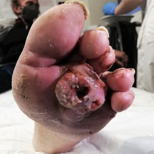
We present here the case of a 55-year-old male patient with a past medical history of HIV infection on appropriate antiretroviral therapy (Bictegravir/Emtricitabine/Tenofovir Alafenamide), previously treated for hepatitis C with sustained viral response, who was diagnosed with squamous cell cancer of his right hand 2 years before, and treated with carboplatin and taxol, and surgery (partial amputation) for cancer progression and recurrent infections. He presented to our emergency department for worsening pain (NRS 10/10) of a suppurative and foul-smelling interdigital ulcerated lesion of his left foot, that had been present for 3 months. He reported 100% adherence to his HIV therapy and denied fever or chills. Initial work-up showed 10,690/mm3 leukocytes (8,240/ mm3 neutrophils, 1,560/mm3 lymphocytes), and a C-reactive protein of 2.89 mg/dL (normal value 0-0.5). CD4 count on presentation was 225 cells/µL. The patient was immediately treated with morphine (10 mg iv) and piperacillin/tazobactam (4.5 gr iv). The lesion presented erythematous ulcerated bleeding surface with serous and fetid secretion on which mobile larvae were found inside (as showed in the video). We removed about 20 maggots with blunt pliers, identified as larvae of Musca domestica (housefly).
Downloads
Villwock JA, Harris TM. Head and neck myiasis, cutaneous malignancy, and infection: A case series and review of the literature. J Emerg Med 2014;47:e37–41. DOI: https://doi.org/10.1016/j.jemermed.2014.04.024
Kondoh A, Ota M, Tokuyama M, et al. Case of Wound Myiasis in a Squamous Cell Carcinoma Lesion of the Scalp. Tokai J Exp Clin Med 2022;47:44-6.
Fukaura R, Terashima-Murase C, Mori S, et al. Myiasis on a spindle cell squamous cell carcinoma: A scanning electron microscope observation of Lucilia sericata larvae [online ahead of print]. J Dermatol 2023;10.1111/1346-8138.16833. DOI: https://doi.org/10.1111/1346-8138.16833
Gonçalves KKN, de Araújo ESM, Barbirato DS, et al. Head and neck cancer associated with myiasis. Int J Oral Maxillofac Surg 2022;51:847-53. DOI: https://doi.org/10.1016/j.ijom.2021.08.011
Jain A. Myiasis in patients with oral squamous cell carcinoma-a systematic review and protocol for management. Oral Maxillofac Surg 2019;23:265-9. DOI: https://doi.org/10.1007/s10006-019-00757-2
Patel BC, Ostwal S, Sanghavi PR, et al. Management of Malignant Wound Myiasis with Ivermectin, Albendazole, and Clindamycin (Triple Therapy) in Advanced Head-and-Neck Cancer Patients: A Prospective Observational Study. Indian J Palliat Care 2018;24:459-64. DOI: https://doi.org/10.4103/IJPC.IJPC_112_18
Waidyaratne G, Zhou S, O'Neil T, Marks A. Management of Wound Myiasis in the Hospice and Palliative Medicine Setting. J Palliat Med 2021;24:797-800. DOI: https://doi.org/10.1089/jpm.2020.0441
Grinblat G, Frenkel Y, Shochat I, et al. Myiasis in Neglected Cutaneous Squamous Cell Carcinoma of the Head and Neck: Review of Management and Current Protocol Recommendations. Adv Skin Wound Care 2021;34:372-8. DOI: https://doi.org/10.1097/01.ASW.0000752708.82300.a4
How to Cite

This work is licensed under a Creative Commons Attribution-NonCommercial 4.0 International License.
PAGEPress has chosen to apply the Creative Commons Attribution NonCommercial 4.0 International License (CC BY-NC 4.0) to all manuscripts to be published.