
Uremic encephalopathy: A definite diagnosis by magnetic resonance imaging?
All claims expressed in this article are solely those of the authors and do not necessarily represent those of their affiliated organizations, or those of the publisher, the editors and the reviewers. Any product that may be evaluated in this article or claim that may be made by its manufacturer is not guaranteed or endorsed by the publisher.
Published: 12 August 2022
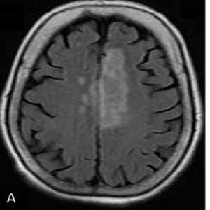
The aim of this study was to investigate the magnetic resonance imaging (MRI) findings for the diagnose uremic encephalopathy and describe the usefulness of MRI findings in the ultimate diagnosis of uremic encephalopathy (UE). A total of 20 patients with uremic encephalopathy admitted to the hospital were evaluated in this prospective study. The clinical manifestations, laboratory and MRI imaging findings, demographic information, and clinical outcome were analyzed for each patient. We observed that the 20 prospectively reviewed patients with UE had no involvement of the basal ganglia or the lentiform fork sign (LFS). However, two-thirds of the patients had white matter involvement, and 80% of the subjects had cerebral or cortical atrophy. The arterial blood gas (ABG) analysis revealed that 50% of the patients suffered from metabolic acidosis (n=10). The results of the present study demonstrated that although the observation of Lentiform Fork Sign and Basal Ganglia involvement in MRI of UE patients is a specific finding the absence of which does not rule out UE. Thus, simultaneous examination of clinical manifestation and laboratory test analyses, along with imaging findings, should also be taken into account.
Downloads
Wang HC, Cheng SJ. The syndrome of acute bilateral basal ganglia lesions in diabetic uremic patients. J Neurol. 2003 Aug;250(8):948-55. DOI: https://doi.org/10.1007/s00415-003-1122-0
Kim DM, Lee IH, Song CJ. Uremic Encephalopathy: MR Imaging Findings and Clinical Correlation. AJNR Am J Neuroradiol. 2016 Sep;37(9):1604-9. Epub 2016 Apr 28. DOI: https://doi.org/10.3174/ajnr.A4776
Lee EJ, Park JH, Ihn Yk, Kim YJ, Lee SK, Park CS. Acute bilateral basal ganglia lesions in diabetic uraemia: diffusion-weighted MRI. Neuroradiology. 2007 Dec;49(12):1009-13. Epub 2007 Oct 6. DOI: https://doi.org/10.1007/s00234-007-0299-9
Yoon CH, Seok JI, Lee DK, An GS. Bilateral basal ganglia and unilateral cortical involvement in a diabetic uremic patient. Clin Neurol Neurosurg. 2009 Jun;111(5):477-9. Epub 2009 Feb 12. DOI: https://doi.org/10.1016/j.clineuro.2009.01.007
Schmidt M, Sitter T, Lederer SR, Held E, Schiffl H. Reversible MRI changes in a patient with uremic encephalopathy. J Nephrol. 2001 Sep-Oct;14(5):424-7.
Tatsumoto N, Fujisaki K, Nagae H, Ono-Fujisaki A, Kura-Nakamura N, Taniguchi M, Masutani K, Tsuruya K, Iida M. Reversible posterior leukoencephalopathy syndrome in a patient with severe uremic encephalopathy. Clin Nephrol. 2010 Aug;74(2):154-8. DOI: https://doi.org/10.5414/CNP74154
Prüss H, Siebert E, Masuhr F. Reversible cytotoxic brain edema and facial weakness in uremic encephalopathy. J Neurol. 2009 Aug;256(8):1372-3. Epub 2009 Apr 12. DOI: https://doi.org/10.1007/s00415-009-5125-3
Kang E, Jeon SJ, Choi SS. Uremic encephalopathy with atypical magnetic resonance features on diffusion-weighted images. Korean J Radiol. 2012 Nov-Dec;13(6):808-11. Epub 2012 Oct 12. DOI: https://doi.org/10.3348/kjr.2012.13.6.808
Casey SO, Sampaio RC, Michel E, Truwit CL. Posterior reversible encephalopathy syndrome: utility of fluid-attenuated inversion recovery MR imaging in the detection of cortical and subcortical lesions. AJNR Am J Neuroradiol. 2000 Aug;21(7):1199-206.
Jarosz JM, Howlett DC, Cox TC, Bingham JB. Cyclosporine-related reversible posterior leukoencephalopathy: MRI. Neuroradiology. 1997 Oct;39(10):711-5. DOI: https://doi.org/10.1007/s002340050492
McKinney AM, Short J, Truwit CL, McKinney ZJ, Kozak OS, SantaCruz KS, Teksam M. Posterior reversible encephalopathy syndrome: incidence of atypical regions of involvement and imaging findings. AJR Am J Roentgenol. 2007 Oct;189(4):904-12. DOI: https://doi.org/10.2214/AJR.07.2024
Mansoor, S, De Klerk L, Lineen J,Fahad M, Ali I, Murphy K Lentiform fork sign in a uremic patient with a high anion gap metabolic acidosis with seizures: a case report from North West of Ireland. The Egyptian Journal of Neurology, Psychiatry and Neurosurgery, 2020. 56(1): 101. DOI: https://doi.org/10.1186/s41983-020-00234-8
Jaryal A, Thakur S, Pathania JS, Vikrant S, Kumar D, Verma L. Lentiform fork sign: Uremia alone or multifactorial causation? Hemodial Int. 2020 Jan;24(1):E10-E12. Epub 2019 Dec 16. DOI: https://doi.org/10.1111/hdi.12810
Kumar G, Goyal MK. Lentiform Fork sign: a unique MRI picture. Is metabolic acidosis responsible? Clin Neurol Neurosurg. 2010 Nov;112(9):805-12. Epub 2010 Jul 7. DOI: https://doi.org/10.1016/j.clineuro.2010.06.006
Grasso D, Borreggine C, Perfetto F, Bertozzi V, Trivisano M, Specchio LM, Grilli G, Macarini L. Lentiform fork sign: a magnetic resonance finding in a case of acute metabolic acidosis. Neuroradiol J. 2014 Jun;27(3):288-92. doi: 10.15274/NRJ-2014-10041. Epub 2014 Jun 17. DOI: https://doi.org/10.15274/NRJ-2014-10041
Fazekas F, Kleinert R, Offenbacher H, Payer F, Schmidt R, Kleinert G, Radner H, Lechner H. The morphologic correlate of incidental punctate white matter hyperintensities on MR images. AJNR Am J Neuroradiol. 1991 Sep-Oct;12(5):915-21.
Pasquier F, Leys D, Weerts JG, Mounier-Vehier F, Barkhof F, Scheltens P. Inter- and intraobserver reproducibility of cerebral atrophy assessment on MRI scans with hemispheric infarcts. Eur Neurol. 1996;36(5):268-72. DOI: https://doi.org/10.1159/000117270
Vanholder R, De Smet R, Glorieux G, Argilés A, Baurmeister U, Brunet P, Clark W, Cohen G, De Deyn PP, Deppisch R, Descamps-Latscha B, Henle T, Jörres A, Lemke HD, Massy ZA, Passlick-Deetjen J, Rodriguez M, Stegmayr B, Stenvinkel P, Tetta C, Wanner C, Zidek W; European Uremic Toxin Work Group (EUTox). Review on uremic toxins: classification, concentration, and interindividual variability. Kidney Int. 2003 May;63(5):1934-43. Erratum in: Kidney Int. 2020 Nov;98(5):1354. DOI: https://doi.org/10.1046/j.1523-1755.2003.00924.x
Thimmaiah R, Murthy KK, Pinto D. Cognitive dysfunction in patients with renal failure requiring hemodialysis. Indian J Psychol Med. 2012;34(3):237-241. DOI: https://doi.org/10.4103/0253-7176.106019
Biasioli S, D'Andrea G, Feriani M, Chiaramonte S, Fabris A, Ronco C, La Greca G. Uremic encephalopathy: an updating. Clin Nephrol. 1986 Feb;25(2):57-63.
Etgen T. Kidney disease as a determinant of cognitive decline and dementia. Alzheimers Res Ther. 2015;7(1):29. Published 2015 Mar 17. DOI: https://doi.org/10.1186/s13195-015-0115-4
Wang HC, Brown P, Lees AJ. Acute movement disorders with bilateral basal ganglia lesions in uremia. Mov Disord. 1998 Nov;13(6):952-7. DOI: https://doi.org/10.1002/mds.870130615
Wali GM, Khanpet MS, Mali RV. Acute movement disorder with bilateral basal ganglia lesions in diabetic uremia. Ann Indian Acad Neurol. 2011 Jul;14(3):211-3. DOI: https://doi.org/10.4103/0972-2327.85899
Albin RL. Basal ganglia neurotoxins. Neurol Clin. 2000 Aug;18(3):665-80. DOI: https://doi.org/10.1016/S0733-8619(05)70217-6
Gong WY, Li SS, Yu ZC, Wu HW, Yin LH, Mei LF, Liu FN. Syndrome of uremic encephalopathy and bilateral basal ganglia lesions in non-diabetic hemodialysis patient: a case report. BMC Nephrol. 2018 Dec 19;19(1):370. DOI: https://doi.org/10.1186/s12882-018-1174-0
Heo K, Cho KH, Lee MK, Chung SJ, Cho YJ, Lee BI. Development of epilepsy after posterior reversible encephalopathy syndrome. Seizure. 2016 Jan;34:90-4. Epub 2015 Dec 18. DOI: https://doi.org/10.1016/j.seizure.2015.12.005
Hobson EV, Craven I, Blank SC. Posterior reversible encephalopathy syndrome: a truly treatable neurologic illness. Perit Dial Int. 2012 Nov-Dec;32(6):590-4. DOI: https://doi.org/10.3747/pdi.2012.00152
Bartynski WS, Boardman JF. Distinct imaging patterns and lesion distribution in posterior reversible encephalopathy syndrome. AJNR Am J Neuroradiol. 2007 Aug;28(7):1320-7. DOI: https://doi.org/10.3174/ajnr.A0549
Kastrup O, Schlamann M, Moenninghoff C, Forsting M, Goericke S. Posterior Reversible Encephalopathy Syndrome: The Spectrum of MR Imaging Patterns. Clin Neuroradiol. 2015 Jun;25(2):161-71. Epub 2014 Feb 20. DOI: https://doi.org/10.1007/s00062-014-0293-7
Johansson BB. The blood-brain barrier and cerebral blood flow in acute hypertension. Acta Med Scand Suppl. 1983;678:107-12. DOI: https://doi.org/10.1111/j.0954-6820.1984.tb08668.x
Tamaki K, Sadoshima S, Baumbach GL, Iadecola C, Reis DJ, Heistad DD. Evidence that disruption of the blood-brain barrier precedes reduction in cerebral blood flow in hypertensive encephalopathy. Hypertension. 1984 Mar-Apr;6(2 Pt 2):I75-81. DOI: https://doi.org/10.1161/01.HYP.6.2_Pt_2.I75
MacKenzie ET, Strandgaard S, Graham DI, Jones JV, Harper AM, Farrar JK. Effects of acutely induced hypertension in cats on pial arteriolar caliber, local cerebral blood flow, and the blood-brain barrier. Circ Res. 1976 Jul;39(1):33-41. DOI: https://doi.org/10.1161/01.RES.39.1.33
Gao B, Yu BX, Li RS, Zhang G, Xie HZ, Liu FL, Lv C. Cytotoxic Edema in Posterior Reversible Encephalopathy Syndrome: Correlation of MRI Features with Serum Albumin Levels. AJNR Am J Neuroradiol. 2015 Oct;36(10):1884-9. Epub 2015 Jul 2. DOI: https://doi.org/10.3174/ajnr.A4379
Casey SO, Truwit CL. Pontine reversible edema: a newly recognized imaging variant of hypertensive encephalopathy? AJNR Am J Neuroradiol. 2000 Feb;21(2):243-5.
De Seze J, Mastain B, Stojkovic T, Ferriby D, Pruvo JP, Destée A, Vermersch P. Unusual MR findings of the brain stem in arterial hypertension. AJNR Am J Neuroradiol. 2000 Feb;21(2):391-4.
Chang GY, Keane JR. Hypertensive brainstem encephalopathy: three cases presenting with severe brainstem edema. Neurology. 1999 Aug 11;53(3):652-4. DOI: https://doi.org/10.1212/WNL.53.3.652
Geboers K, Vanden Bossche S, Dekeyzer S. Lentiform fork sign in a girl with uremic encephalopathy. Acta Neurol Belg. 2022 Apr;122(2):535-536. Epub 2021 Aug 3. DOI: https://doi.org/10.1007/s13760-021-01764-5
Soung-Rok SIM, Sang-Hun LEE, Jae-Hoon JAHNG, Jae-Yun LIM, You-Kyoung CHOI, Ki-Sun BAE, Woo-Il PARK, Ki-Joong KIM, Kyung-Yul LEE, Hyeong-Cheon PARK, Sung-Kyu HA. Uremic encephalopathy associated with bilateral basal ganglia and cerebellar lesion in a non-diabetic hemodialysis patient. Korean Journal of Nephrology, 2006. 25(6): 1061-1066.
Sharrief AZ, Raffel J, Zee DS. Vitamin B(12) deficiency with bilateral globus pallidus abnormalities. Arch Neurol. 2012 Jun;69(6):769-772. DOI: https://doi.org/10.1001/archneurol.2011.1084
Hassanzadeh S, Jalessi M, Jameie SB, Khanmohammadi M, Bagher Z,Namjoo, Z, Davachi SM. More attention on glial cells to have better recovery after spinal cord injury. Biochemistry and Biophysics Reports, 2021. 25:100905. DOI: https://doi.org/10.1016/j.bbrep.2020.100905
How to Cite
PAGEPress has chosen to apply the Creative Commons Attribution NonCommercial 4.0 International License (CC BY-NC 4.0) to all manuscripts to be published.