
Length-dependent changes of lower limb muscle morphology in Chronic Inflammatory Demyelinating Polyneuropathy assessed with magnetic resonance imaging
All claims expressed in this article are solely those of the authors and do not necessarily represent those of their affiliated organizations, or those of the publisher, the editors and the reviewers. Any product that may be evaluated in this article or claim that may be made by its manufacturer is not guaranteed or endorsed by the publisher.
Published: 18 November 2021
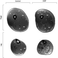
The objective of the present study was to assess muscle quantity of the thigh and leg in patients with chronic inflammatory demyelinating polyneuropathy (CIDP) compared to age and sex matched controls in exploring length-dependent changes of innervated muscles. In five people with CIDP and seven controls, magnetic resonance imaging was used to assess muscle morphology of the four parts of the quadriceps and medial hamstring muscles. Findings were compared to the triceps surae from a subset of participants. The CIDP group had less contractile tissue in the quadriceps (11.5%, P<0.05), hamstrings (15.6%, P<0.05) and triceps surae (35.9%, P<0.05) compared to controls. Additionally, CIDP had less contractile tissue (18.7%) in the triceps surae compared to the hamstrings (P<0.05). Muscle quantity in the quadriceps and hamstrings in CIDP was less than controls, but differences were greater for the distal triceps surae. These findings support a length-dependent affect of CIDP on limb musculature composition.
Downloads
Bromberg M. What is in the literature. J Clin Neuromuscul Dis. 2021 Jun 1;22(4):200-208. DOI: https://doi.org/10.1097/CND.0000000000000371
Bosboom WMJ, van den Berg LH, Franssen H, Giesbergen PCLM, Flach HZ, van Putten AM, Veldman H, Wokke HJ. Diagnostic value of sural nerve demyelination in chronic inflammatory demyelinating polyneuropathy. Brain. 2001 Dec;124(Pt 12):2427-38. DOI: https://doi.org/10.1093/brain/124.12.2427
Piccolino M. Luigi Galvani's path to animal electricity. J Hist Neurosci. 2008;17(3):335-48. DOI: https://doi.org/10.1080/09647040701420198
Seyfarth EA. Julius Bernstein (1839-1917): pioneer neurobiologist and biophysicist. Biol Cybern. 2006 Jan;94(1):2-8. DOI: https://doi.org/10.1007/s00422-005-0031-y
Shah S, Morrow JM, Sinclair CDJ, Reilly MM, Thornton JS, Lunn MP, Yousry TA. MRI quantifies lumbosacral nerve root and sciatic nerve hypertrophy in chronic inflammatory demyelinating polyradiculoneuropathy. Eur J Radiol. 2020 Sep;130:109164. DOI: https://doi.org/10.1016/j.ejrad.2020.109164
Pitarokoili K, Schlamann M, Kerasnoudis A, Gold R, Yoon M-S. Comparison of clinical, electrophysiological, sonographic and MRI features in CIDP. J Neurol Sci. 2015 Oct 15;357(1-2):198-203. DOI: https://doi.org/10.1016/j.jns.2015.07.030
Ishikawa T, Asakura K, Mizutani Y, Ueda A, Murate K-I, Hikichi C, et al. Magnetic resonance neurography for the evaluation of CIDP. Muscle Nerve. 2017 Apr;55(4):483-489. DOI: https://doi.org/10.1002/mus.25368
Sinclair CDJ, Morrow JM, Miranda MA, Davagnanam I, Cowley PC, Mehta H, Hanna M, Koltzenburg M, Yousry T, Reilly M, Thornton JS. Skeletal muscle MRI magnetisation transfer ratio reflects clinical severity in peripheral neuropathies. J Neurol Neurosurg Psychiatry. 2012 Jan;83(1):29-32. DOI: https://doi.org/10.1136/jnnp.2011.246116
Gilmore KJ, Doherty TJ, Kimpinski K, Rice CL. Reductions in muscle quality and quantity in chronic inflammatory demyelinating polyneuropathy patients assessed by magnetic resonance imaging. Muscle Nerve. 2018 Sep;58(3):396-401. DOI: https://doi.org/10.1002/mus.26159
Gilmore KJ, Fanous J, Doherty TJ, Kimpinski K, Rice CL. Nerve dysfunction leads to muscle morphological abnormalities in chronic inflammatory demyelinating polyneuropathy assessed by MRI. Clin Anat. 2020 Jan;33(1):77-84. DOI: https://doi.org/10.1002/ca.23473
Harbo T, Andersen H, Jakobsen J. Length-dependent weakness and electrophysiological signs of secondary axonal loss in chronic inflammatory demyelinating polyradiculoneuropathy. Muscle Nerve. 2008 Aug;38(2):1036-45. DOI: https://doi.org/10.1002/mus.21000
Bergh FPYK Van Den, Hadden RDM, Bouche P. European Federation of Neurological Societies / Peripheral Nerve Society Guideline on management of chronic inflammatory demyelinating polyradiculoneuropathy: Report of a joint task force of the European Federation of Neurological Societies and the Peripheral Nerve Society - First Revision. Eur J Neurol. 2010 Mar;17(3):356-63. DOI: https://doi.org/10.1111/j.1468-1331.2009.02930.x
Ohyama K, Koike H, Katsuno M, Takahashi M, Hashimoto R, Kawagashira Y, et al. Muscle atrophy in chronic inflammatory demyelinating polyneuropathy: A computed tomography assessment. Eur J Neurol. 2014 Jul;21(7):1002-10. DOI: https://doi.org/10.1111/ene.12426
Fisse AL, Fiegert S, Stoykova Z, Brunger J, Athanasopoulos D, Gruter T, Motte J, Gold R, Pitarokoili K. Increased muscle echointensity correlates with clinical disability and muscle strength in chronic inflammatory demyelinating polyneuropathy. Eur J Neurol. 2021 May;28(5):1698-1705. DOI: https://doi.org/10.1111/ene.14716
Kuwabara S, Ogawara K, Misawa S, Mori M, Hattori T. Distribution patterns of demyelination correlate with clinical profiles in chronic inflammatory demyelinating polyneuropathy. J Neurol Neurosurg Psychiatry. 2002 Jan;72(1):37-42. DOI: https://doi.org/10.1136/jnnp.72.1.37
Kuwabara S, Misawa S. Chronic inflammatory demyelinating polyneuropathy: Clinical subtypes and their correlation with electrophysiology. Clinical & Experimental Neuroimmunology. 2011;2:41-48. DOI: https://doi.org/10.1111/j.1759-1961.2011.00020.x
Abe T, Loenneke JP, Thiebaud RS, Loftin M. Age-related muscle loss of the anterior and posterior thigh assessed by means of MRI/CT and ultrasound. Journal of Trainology. 2014; 3:47-52. DOI: https://doi.org/10.17338/trainology.3.2_47
Kilroe SP, Fulford J, Jackman SR, Vanloon LJC, Wall BT. Temporal muscle-specific disuse atrophy during one week of leg immobilization. Med Sci Sports Exerc. 2020 Apr;52(4):944-954. DOI: https://doi.org/10.1249/MSS.0000000000002200
Maden-Wilkinson T, Degens H, Jones DA, McPhee JS. Comparison of MRI and DXA to measure muscle size and age-related atrophy in thigh muscles. J Musculoskelet Neuronal Interact. 2013 Sep;13(3):320-8.
Hogrel JY, Barnouin Y, Azzabou N, Butler-Browne G, Voit T, Moraux A, Leroux G, Behin A, McPhee JS, Carlier PG. NMR imaging estimates of muscle volume and intramuscular fat infiltration in the thigh: variations with muscle, gender, and age. Age (Dordr). 2015 Jun;37(3):9798. DOI: https://doi.org/10.1007/s11357-015-9798-5
Morse CI, Thom JM, Reeves ND, Birch KM, Narici MV. In vivo physiological cross-sectional area and specific force are reduced in the gastrocnemius of elderly men. J Appl Physiol (1985). 2005 Sep;99(3):1050-5. DOI: https://doi.org/10.1152/japplphysiol.01186.2004
Ginsberg L, Platts AD, Thomas PK. Chronic inflammatory demyelinating polyneuropathy mimicking a lumbar spinal stenosis syndrome. J Neurol Neurosurg Psychiatry. 1995 Aug;59(2):189-91. DOI: https://doi.org/10.1136/jnnp.59.2.189
Piccione EA, Engelstad J, Dyck PJ, Mauremann ML, Dispenzieri A, Dyck JB. Nerve pathologic features differentiate POEMS syndrome from CIDP. Acta Neuropathol Commun. 2016 Oct 31;4(1):116. DOI: https://doi.org/10.1186/s40478-016-0389-1
Dahlqvist JR, Salim R, Thomsen C, Vissing J. A quantitative method to assess muscle edema using short T1 inversion recovery MRI. Sci Rep. 2020 Apr 29;10(1):7246. DOI: https://doi.org/10.1038/s41598-020-64287-8
Edmunds KJ, Okonkwo OC, Sigurdsson S, Lose SR, Gudnason V, Carraro U, Gargiulo P. Soft tissue radiodensity parameters mediate the relationship between self-reported physical activity and lower extremity function in AGES-Reykjavík participants. Sci Rep. 2021 Oct 11;11(1):20173. DOI: https://doi.org/10.1038/s41598-021-99699-7
Ricciardi C, Edmunds KJ, Recenti M, Sigurdsson S, Gudnason V, Carraro U, Gargiulo P. Assessing cardiovascular risks from a mid-thigh CT image: a tree-based machine learning approach using radiodensitometric distributions. Sci Rep. 2020 Feb 18;10(1):2863. DOI: https://doi.org/10.1038/s41598-020-59873-9
Recenti M, Ricciardi C, Edmunds K, Gislason MK, Gargiulo P. Machine learning predictive system based upon radiodensitometric distributions from mid-thigh CT images. Eur J Transl Myol. 2020 Apr 1;30(1):8892. eCollection 2020 Apr 7. DOI: https://doi.org/10.4081/ejtm.2019.8892
How to Cite
PAGEPress has chosen to apply the Creative Commons Attribution NonCommercial 4.0 International License (CC BY-NC 4.0) to all manuscripts to be published.