
Ovine fetuses from slaughterhouses: A useful source for neural cell primary cultures
All claims expressed in this article are solely those of the authors and do not necessarily represent those of their affiliated organizations, or those of the publisher, the editors and the reviewers. Any product that may be evaluated in this article or claim that may be made by its manufacturer is not guaranteed or endorsed by the publisher.
Published: 22 April 2021
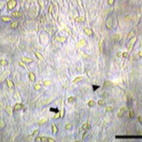
A lot of evidence demonstrates that sheep could represent an experimental model to set up medical procedures in view of their application on humans. Sheep are chosen as models for human biomechanical studies because their skeleton has some similarities to humans. The aim of this work was to set up sheep primary cultures from ovine fetuses at different ages, from pregnant uteri retrieved at local abattoirs. Cell characterization showed that one cell population was immunopositive to GFAP and identifiable as astrocytes, whereas a second cell type was III β-tubulin-positive, and hence classified as neurons. At 60-days old fetus is suitable to obtain neurons, whereas in a 90-days old fetus the cell culture is predominantly characterized by glial cells. The procedure here proposed is inexpensive, in fact, collecting fetuses during sheep slaughtering is a cost-saving option, unlike common experimental animals such as mice, rats, rabbits, that require very high economical efforts. Finally, our protocol fully eliminates the need of animal killing, being living animals replaced by a validated in vitro model in agreement with the 3Rs statement.
Downloads
Hashimoto A, Onodera T, Ikeda H, Kitani H. Isolation and characterization of fetal bovine brain cells in primary culture. Res Vet Sci 2000;69:39-46. DOI: https://doi.org/10.1053/rvsc.2000.0382
Peruffo A, Cozzi B. Bovine brain: an in vitro translational model in developmental neuroscience and neurodegenerative research. Front in Pediatr 2014;2:74. DOI: https://doi.org/10.3389/fped.2014.00074
Gadau SD. Tubulin post-translational modifications in developing dog primary neurons obtained with methods according to the 3Rs principles. Res Vet Sci 2019;122:56-63. DOI: https://doi.org/10.1016/j.rvsc.2018.11.015
Martini L, Fini M, Giavaresi G, Giardino R. Sheep model in orthopedic research: a literature review. Comp Med 2001;51:292-9.
Holt JD, Cameron D, Dias N, et al. The sheep as a model of preclinical safety and pharmacokinetic evaluations of candidate microbicides. Antimicrob Agents Chemother 2015;59:3761-70. DOI: https://doi.org/10.1128/AAC.04954-14
Braak H, Braak E, Strothjohann M. Abnormally phosphorylated tau protein related to the formation of neurofibrillary tangles and neuropil threads in the cerebral cortex of sheep and goat. Neurosci Lett 1994;171:1-4. DOI: https://doi.org/10.1016/0304-3940(94)90589-4
Van Dam D, De Deyn PP. Animal models in the drug discovery pipeline for Alzheimer's disease. Br J Pharmacol 2011;164:1285-300. DOI: https://doi.org/10.1111/j.1476-5381.2011.01299.x
Kay GW, Oswald MJ, Palmer DN. The development and characterisation of complex ovine neuron cultures from fresh and frozen foetal neurons. J Neurosci Methods 2006;155:98-108. DOI: https://doi.org/10.1016/j.jneumeth.2006.01.008
Roselli CE, Larkin K, Resko JA, et al. The volume of a sexually dimorphic nucleus in the ovine medial preoptic area/anterior hypothalamus varies with sexual partner preference. Endocrinology 2004;145:478-83. DOI: https://doi.org/10.1210/en.2003-1098
Mura A, Gadau S, Lepore G, et al. Expression and distribution of P450-aromatase in the ovine hypothalamus at different stages of fetal development. Neuro Endocrinol Lett 2010;31:690-9.
Lepore G, Gadau S, Mura A, et al. Aromatase immunoreactivity in fetal ovine neuronal cell cultures exposed to oxidative injury. Eur J Histochem 2009;53:233-8. DOI: https://doi.org/10.4081/ejh.2009.233
Lepore G, Gadau S, Peruffo A, et al. Aromatase expression in cultured fetal sheep astrocytes after nitrosative/oxidative damage. Cell Tissue Res 2011;344:407-13. DOI: https://doi.org/10.1007/s00441-011-1160-3
Lepore G, Zedda M, Mura E, et al. Brain aging and testosterone-induced neuroprotection: studies on cultured sheep cortical neurons. Neuro Endocrinol Lett 2013;34:395-401.
Mura E, Lepore G, Zedda M, et al. Sheep primary astrocytes under starvation conditions express higher amount of LC3 II autophagy marker than neurons. Arch Ital Biol 2014;152:47-56.
Farina V, Lepore G, Biagi F, et al. Autophagic processes increase during senescence in cultured sheep neurons and astrocytes. Eur J Histochem 2018;62:22-7. DOI: https://doi.org/10.4081/ejh.2018.2891
Hill E, Nagel D, Parri R, Coleman M. Stem cell-derived astrocytes: are they physiologically credible? J Physiol 2016;594:6595-606. DOI: https://doi.org/10.1113/JP270658
Grainger AI, King MC, Nagel DA, et al. In vitro Models for Seizure-Liability Testing Using Induced Pluripotent Stem Cells. Front Neurosci 2018;12:590. DOI: https://doi.org/10.3389/fnins.2018.00590
Logan S, Arzua T, Canfield SG, et al. Studying Human Neurological Disorders Using Induced Pluripotent Stem Cells: From 2D Monolayer to 3D Organoid and Blood Brain Barrier Models. Compar Physiol 2019;9:565-611. DOI: https://doi.org/10.1002/cphy.c180025
Polin RA, Abman SH, Rowitch D, Benitz WE. Fetal and Neonatal Physiology. New York: Elsevier; 2016.
McGeady TA, Quinn PJ, Fitzpatrick ES, Ryan MT. Veterinary Embryology. Blackwell, Oxford 2006.
Gadau SD. Morphological and quantitative analysis on a-tubulin modifications in glioblastoma cells. Neurosci Lett 2018. DOI: https://doi.org/10.1016/j.neulet.2018.09.044
Dráberová E, Lukás Z, Ivanyi D, et al. Expression of class III beta-tubulin in normal and neoplastic human tissues. Histochem Cell Biol 1998;109:231-9. DOI: https://doi.org/10.1007/s004180050222
Schock SC, Jolin-Dahel KS, Schock PC, et al. Striatal interneurons in dissociated cell culture. Histochem Cell Biol 2010;134:1-12. DOI: https://doi.org/10.1007/s00418-010-0707-9
Hofman MA. Size and shape of the cerebral cortex in mammals. The cortical surface. Brain Behav Evol 1985;27:28-40. DOI: https://doi.org/10.1159/000118718
Gibbons HM, Dragunow M. Adult human brain cell culture for neuroscience research. The Int J Biochem Cell Biol 2010;2:844-56. DOI: https://doi.org/10.1016/j.biocel.2009.12.002
Townes-Anderson E, MacLeish PR, Raviola E. Rod cells dissociated from mature salamander retina: ultrastructure and uptake of horseradish peroxidase. J Cell Biol 1985;100:175-88. DOI: https://doi.org/10.1083/jcb.100.1.175
Bologa L, Joubert R, Bisconte JC, et al. Development of immunologically identified brain cells in culture: quantitative aspects. Exp Brain Res 1983;53:163-7. DOI: https://doi.org/10.1007/BF00239408
Ranganatha N, Kuppast IJ. A review on alternatives to animal testing methods in drug development. Int J Pharm Pharmaceut Sci 2012;4:28-32.
Doke SK, Dhawale SC. Alternatives to animal testing: A review. Saudi Pharm J 2015;23:223-9. DOI: https://doi.org/10.1016/j.jsps.2013.11.002
Price DL, Sisodia SS, Koo EH, et al. Neuronal disorders: studies of animal models and human diseases. Toxicol Pathol 1990;18:128-37. DOI: https://doi.org/10.1177/019262339001800118
How to Cite
PAGEPress has chosen to apply the Creative Commons Attribution NonCommercial 4.0 International License (CC BY-NC 4.0) to all manuscripts to be published.