
Can MRI quantify the volume changes of denervated facial muscles?
All claims expressed in this article are solely those of the authors and do not necessarily represent those of their affiliated organizations, or those of the publisher, the editors and the reviewers. Any product that may be evaluated in this article or claim that may be made by its manufacturer is not guaranteed or endorsed by the publisher.
Published: 1 April 2020
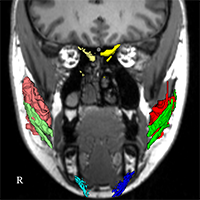
Could manual segmentation of magnetic resonance images be used to quantify the effects of transcutaneous electrostimulation and reinnervation of denervated facial muscle? Five patients with unilateral facial paralysis were scanned during the study while receiving a daily surface electrostimulation of the paralytic cheek region, but also after reinnervation. Their facial muscles were identified in 3D (coronal, sagittal, and axial) and segmented in magnetic resonance imaging (MRI) data for in total 28 time points over the 12 months of study. A non-significant trend of increasing muscle volume were detected after reinnervation. MRI is a valuable technique in the facial paralysis research.
Downloads
Stuart RM, Byrne PJ. The importance of facial expression and the management of facial nerve injury. Neurosurg Q. 2004. doi:10.1097/00013414-200412000-00009 DOI: https://doi.org/10.1097/00013414-200412000-00009
Coulson SE, O’dwyer NJ, Adams RD, Croxson GR. Expression of emotion and quality of life after facial nerve paralysis. Otol Neurotol Off Publ Am Otol Soc Am Neurotol Soc [and] Eur Acad Otol Neurotol. 2004;25(6):1014-1019. doi:10.1097/00129492-200411000-00026 DOI: https://doi.org/10.1097/00129492-200411000-00026
Butler DP, Grobbelaar AO. Facial palsy: what can the multidisciplinary team do? J Multidiscip Healthc. 2017;10:377-381. doi:10.2147/JMDH.S125574 DOI: https://doi.org/10.2147/JMDH.S125574
Harrison DH, Grobbelaar AO. Pectoralis minor muscle transfer for unilateral facial palsy reanimation: An experience of 35 years and 637 cases. J Plast Reconstr Aesthetic Surg. 2012;65(7):845-850. doi:10.1016/j.bjps.2012.01.024 DOI: https://doi.org/10.1016/j.bjps.2012.01.024
Guntinas-Lichius O, Glowka TR, Angelov DN, Irintchev A, Neiss WF. Improved functional recovery after facial nerve reconstruction by temporary denervation of the contralateral mimic musculature with botulinum toxin in rats. Neurorehabil Neural Repair. 2011;25(1):15-23. doi:10.1177/1545968310376058 DOI: https://doi.org/10.1177/1545968310376058
Targan RS, Alon G, Kay SL. Effect of long-term electrical stimulation on motor recovery and improvement of clinical residuals in patients with unresolved facial nerve palsy. Otolaryngol Head Neck Surg. 2000;122(2):246-252. doi:10.1016/S0194-5998(00)70248-8 DOI: https://doi.org/10.1016/S0194-5998(00)70248-8
Teixeira LJ, Valbuza JS, Prado GF. Physical therapy for Bell’s palsy (idiopathic facial paralysis). Cochrane database Syst Rev. 2011;(12):CD006283. doi:10.1002/14651858.CD006283.pub3 DOI: https://doi.org/10.1002/14651858.CD006283.pub3
Kern H, Carraro U, Adami N, et al. One year of home-based daily FES in complete ower motor neuron paraplegia: Recovery of etanic contractility drives the structural mprovements of denervated muscle. Neurol Res. 2010;32:5-12. doi:10.1179/174313209X385644 DOI: https://doi.org/10.1179/174313209X385644
Willand MP. Electrical Stimulation Enhances Reinnervation After Nerve Injury. Eur J Transl Myol. 2015;25(4):243-248. doi:10.4081/ejtm.2015.5243 DOI: https://doi.org/10.4081/ejtm.2015.5243
Shapira Y, Sammons V, Forden J, et al. Brief Electrical Stimulation Promotes Nerve Regeneration Following Experimental In-Continuity Nerve Injury. Neurosurgery. 2018;85(1):156-163. doi:10.1093/neuros/nyy221 DOI: https://doi.org/10.1093/neuros/nyy221
Bueno CR de S, Pereira M, Favaretto IAJ, et al. Electrical stimulation attenuates morphological alterations and prevents atrophy of the denervated cranial tibial muscle. Einstein (Sao Paulo). 2017;15(1):71-76. doi:10.1590/S1679-45082017AO3808 DOI: https://doi.org/10.1590/s1679-45082017ao3808
Kern H, Gargiulo P, Pond A, Albertin G, Marcante A, Carraro U. To Reverse Atrophy of Human Muscles in Complete SCI Lower Motor Neuron Denervation by Home-Based Functional Electrical Stimulation. Adv Exp Med Biol. 2018;1088:585-591. doi:10.1007/978-981-13-1435-3_27 DOI: https://doi.org/10.1007/978-981-13-1435-3_27
Puls WC, Jarvis JC, Ruck A, Lehmann T, Guntinas-Lichius O, Volk GF. Surface electrical stimulation for facial paralysis is not harmful. Muscle Nerve. 2020;61(3):347-353. doi:10.1002/mus.26784 DOI: https://doi.org/10.1002/mus.26784
Volk GF, Thielker J, Moller MC, et al. Tolerability of facial electrostimulation in healthy adults and patients with facial synkinesis. Eur Arch Otorhinolaryngol. January 2020. doi:10.1007/s00405-020-05818-x DOI: https://doi.org/10.1007/s00405-020-05818-x
Grosheva M, Guntinas-Lichius O. Significance of electromyography to predict and evaluate facial function outcome after acute peripheral facial palsy. Eur Arch Otorhinolaryngol. 2007;264(12):1491-1495. doi:10.1007/s00405-007-0376-z DOI: https://doi.org/10.1007/s00405-007-0376-z
Mugler III JP, Brookeman JR. Rapid three-dimensional T1-weighted MR imaging with the MP-RAGE sequence. J Magn Reson Imaging. 1991;1(5):561-567. doi:10.1002/jmri.1880010509 DOI: https://doi.org/10.1002/jmri.1880010509
Song Y, Forsgren S, Yu J, Lorentzon R, Stål PS. Effects on contralateral muscles after unilateral electrical muscle stimulation and exercise. PLoS One. 2012;7(12):e52230-e52230. doi:10.1371/journal.pone.0052230 DOI: https://doi.org/10.1371/journal.pone.0052230
Engelke K, Museyko O, Wang L, Laredo J-D. Quantitative analysis of skeletal muscle by computed tomography imaging-State of the art. J Orthop Transl. 2018;15:91-103. doi:10.1016/j.jot.2018.10.004 DOI: https://doi.org/10.1016/j.jot.2018.10.004
Burmeister H, Baltzer P, Volk G, et al. Evaluation of the early phase of Bell’s palsy using 3 T MRI. Eur Arch Otorhinolaryngol. 2011;268:1493-1500. doi:10.1007/s00405-011-1498-x DOI: https://doi.org/10.1007/s00405-011-1498-x
Volk GF, Karamyan I, Klingner CM, Reichenbach JR, Guntinas-Lichius O. Quantitative magnetic resonance imaging volumetry of facial muscles in healthy patients with facial palsy. Plast Reconstr surgery Glob open. 2014;2(6):e173. doi:10.1097/GOX.0000000000000128 DOI: https://doi.org/10.1097/GOX.0000000000000128
Gargiulo P, Klingner C, Fridgeirsson E, Burmeister H, Volk G, Guntinas-Lichius O. Side differences in MRI-scans in facial palsy: 3-D modeling, segmentation and gray value analysis. 23rd Eur Model Simul Symp EMSS 2011. January 2011:87-92.
How to Cite
PAGEPress has chosen to apply the Creative Commons Attribution NonCommercial 4.0 International License (CC BY-NC 4.0) to all manuscripts to be published.