

 https://doi.org/10.4081/ecj.2026.15940
07 | Dizziness that makes the emergency physician tremble
All claims expressed in this article are solely those of the authors and do not necessarily represent those of their affiliated organizations, or those of the publisher, the editors and the reviewers. Any product that may be evaluated in this article or claim that may be made by its manufacturer is not guaranteed or endorsed by the publisher.
Published: 15 July 2026
Background. Abdominal Aortic Aneurysm (AAA) is amongst the conditions that most concern Emergency Department (ED) physicians, as its diagnosis is often delayed and its rupture carries a very high mortality risk. It is often such a sudden and devastating event that patients may not even make it to the nearest hospital, making it one of the deadliest conditions, with a mortality rate around 90%. Onset is often sudden, but in some cases the rupture is minor or contained, and the patient may present with poorly localized abdominal pain, lower back pain, a palpable abdominal mass, or shock characterized by clammy skin and tachycardia.
Case Presentation. Patient male, 74 years old, hypertension, former smoker, ischemic heart disease with PM, diabetes mellitus, thyroid dysfunction.
Presentation to the ED shock room (Barletta Hospital): “Episode of dizziness with loss of sphincter control, and pain in the left chest and left flank, ongoing for two days.”
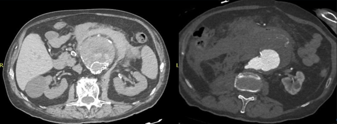
The patient arrived at the ED autonomously and was admitted to the shock room within minutes. Vital signs were within normal limits, GCS 15, troponin 6.5 ng/L, ABG showed mild hypoxia with oxygen saturation at 94%, lactate 1.15 mmol/L, and hemoglobin 12.7 g/dL. He appeared pale, tachycardic, and diaphoretic. A POCUS abdominal scan was performed, immediately revealing a large, round mass located in the left hypochondrium and mesogastrium. Suspicion of a dissection was raised, later confirmed by CT scan. Two peripheral IV lines were placed, and the patient was promptly treated with: IV fluids, Tranexamic acid: 1 g bolus + two slow vials, Andexanet and coagulation factors, Sedoanalgesia with morphine. Due to a hemoglobin drop of about 3 g/dL, infusion of two units of packed red blood cells was started. The patient remained hemodynamically stable and was transferred with ICU-level transport to Vascular Surgery, where he underwent surgery and was discharged five days later.
Conclusions. In the presented case the key to a favorable outcome was the triage nurse's recognition of the patient as high-risk, allowing for immediate access to care. The execution of the E-FAST was crucial, as it enabled an early diagnostic suspicion, leading to the positive outcome for the patient.
Downloads
-
CRediT authorship contribution
Supporting Agencies
Data Availability Statement
How to Cite

This work is licensed under a Creative Commons Attribution-NonCommercial 4.0 International License.
PAGEPress has chosen to apply the Creative Commons Attribution NonCommercial 4.0 International License (CC BY-NC 4.0) to all manuscripts to be published.