Skip to main content Skip to main navigation menu Skip to site footer
Microscopie

  • Archives
  • Login
This journal has not published any issues.
  1. Home /
  2. Search

Search

Advanced filters
Published After
Published Before

Search Results

##search.searchResults.foundPlural##
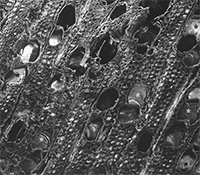
  • Scanning and transmission electron microscopy in cultural heritage: State of the art

    Sabrina Burattini, Elisabetta Falcieri
    23-06-2020
    https://doi.org/10.4081/microscopie.2020.9183
    1198
    Downloads: 592
  • Diaminobenzidine photoconversion allows detection of fluorescently-labelled nanoparticles at transmission electron microscopy after embedding in epoxy and acrylic resins

    B. Cisterna, M. Costanzo, M. Giagnacovo, V. Galimberti, M. Malatesta, C. Zancanaro
    45-49
    30-09-2012
    https://doi.org/10.4081/microscopie.2012.4984
    817
    Downloads: 1491
  • Study of the asbestos bodies and chemical-physical modification of mineral fibres in rat histological tissues using scanning electron microscopy, and high-resolution transmission electron microscopy

    Nicola Bursi Gandolfi
    29-03-2018
    https://doi.org/10.4081/microscopie.2018.7321
    944
    Downloads: 557
  • Ultrastructural immunolocalization of the senescence-related protein terminin in human fibroblasts

    M. Giagnacovo, M. Malatesta, P. Veneroni, E. Wang, C. Pellicciari
    32-37
    30-09-2010
    https://doi.org/10.4081/microscopie.2010.4972
    586
    Downloads: 918
  • Microscopic evaluation of the enamel surface after debonding procedures: An ex vivo study using scanning electron microscopy

    Sara Bernardi, Maria A. Continenza, Guido Macchiarelli
    29-03-2018
    https://doi.org/10.4081/microscopie.2018.7190
    1257
    Downloads: 857
  • Scanning electron microscopy in the taxonomical study of free-living marine nematodes

    Lucia Cesaroni, Loretta Guidi, Maria Balsamo, Federica Semprucci
    31-38
    27-10-2017
    https://doi.org/10.4081/microscopie.2017.6970
    1928
    Downloads: 1558
  • New insight into the information carried by electrons

    Federico Venturi
    19-10-2018
    https://doi.org/10.4081/microscopie.2018.7727
    566
    Downloads: 308
  • The contribution of microscopy to food science: State of the art

    Manuela Malatesta
    19-10-2018
    https://doi.org/10.4081/microscopie.2018.7807
    1251
    Downloads: 530
  • Microscopy techniques in nanomedical research

    Manuela Costanzo, Flavia Carton, Manuela Malatesta
    66-71
    29-03-2017
    https://doi.org/10.4081/microscopie.2017.6732
    1086
    Downloads: 1559
  • Myofibrillar myopathies through the microscope: From diagnosis to molecular pathogenesis

    Valeria Guglielmi, Giuliano Tomelleri, Gaetano Vattemi
    29-03-2018
    https://doi.org/10.4081/microscopie.2018.7325
    1355
    Downloads: 991
  • Melatonin prevents cisplatin- and staurosporine-induced apoptosis in skeletal muscle cells in vitro

    Valentina Baldassarri, Barbara Canonico, Sabrina Burattini, Michela Battistelli, Stefano Papa, Elisabetta Falcieri, Sara Salucci
    19-10-2018
    https://doi.org/10.4081/microscopie.2018.7334
    685
    Downloads: 353
  • Contribution of light and electron microscopy in the identification of morphological alterations in large Japanese field mouse (Apodemus speciosus) testes exposed to low-dose-rate radiations

    Marta Gatti, Maria Grazia Palmerini, Manuel Belli, Stefania Annarita Nottola, Yohei Fujishima, Hideaki Yamashiro, Akifumi Nakata, Tomosato Miura, Guido Macchiarelli
    02-03-2021
    https://doi.org/10.4081/microscopie.2020.9697
    942
    Downloads: 366
  • Ultrastructure of pasta from gluten-free grains: a brief overview

    Mattia Grumi, Paolo Maciotta, Paolo D'Incecco, Alessandra Marti
    28-06-2021
    https://doi.org/10.4081/microscopie.2021.9884
    1951
    Downloads: 572
  • In vitro ageing of myotubes derived from myoblasts of patients affected by myotonic dystrophy type 2: ultrastructural evidence

    M. Giagnacovo, M. Costanzo, R. Cardani, P. Veneroni, C. Pellicciari, G. Meola
    60-67
    31-03-2011
    https://doi.org/10.4081/microscopie.2011.4978
    605
    Downloads: 1138
  • Scanning electron microscopy in monitoring the aging of alternative materials for plastering of canvas manufactured products

    S. Burattini, L. Baratin, L. Borgioli, L. Sabatini, S. Orsini, V. Viti, E. Falcieri, D. De Luca
    47-51
    31-03-2014
    https://doi.org/10.4081/microscopie.2014.4990
    755
    Downloads: 1545
  • Imaging in nanomedicine: a multidisciplinary challenge

    Manuela Malatesta
    28-09-2020
    https://doi.org/10.4081/microscopie.2020.9343
    568
    Downloads: 409
  • Electron microscopy to characterise nanoparticles in the biological environment

    Mathieu Repellin, Flavia Carton
    01-04-2019
    https://doi.org/10.4081/microscopie.2019.8188
    1011
    Downloads: 407
  • A metallographic approach to the study of MgH2-Mg phase transformation

    M. Vittori Antisari, A. Montone, N. Abazovic, A. Aurora, M. Drvendzija, M.R. Mancini, D. Mirabile Gattia, F. Pierdominici
    53-57
    31-03-2008
    https://doi.org/10.4081/microscopie.2008.4959
    833
    Downloads: 1157
  • Functional nanostructures and interfaces of strontium ruthenates Sr3Ru2O7/Sr2RuO4 eutectic crystals

    R. Ciancio, H. Pettersson, J. Börjesson, S. Lopatin, R. Fittipaldi, A. Vecchione, S. Kittaka, Y. Maeno, S. Pace, E. Olsson
    36-43
    31-03-2010
    https://doi.org/10.4081/microscopie.2010.4968
    655
    Downloads: 1250
  • TEM investigation of the crystal lattice registration of carbon nanotubes over graphene membranes

    L. Ortolani, F. Houdellier, M. Monthioux, V. Morandi
    43-49
    30-09-2010
    https://doi.org/10.4081/microscopie.2010.4974
    940
    Downloads: 1249
  • Ultrastructural characterization of the MBNL1- containing foci occurring in myoblast and myotube nuclei from patients affected by myotonic dystrophy type 2

    F. Perdoni, R. Cardani, M. Giagnacovo, P. Veneroni, G. Meola, C. Pellicciari, M. Malatesta
    60-64
    30-09-2009
    https://doi.org/10.4081/microscopie.2009.4967
    487
    Downloads: 955
  • Ultrastructural study of the main epigenetic markers in hepatocyte nuclei

    Maria Assunta Lacavalla, Stella Siciliani
    07-04-2020
    https://doi.org/10.4081/microscopie.2020.8996
    620
    Downloads: 347
  • Atomic structure and crystallographic shear planes in epitaxial TiO2 anatase thin films

    R. Ciancio, A. Vittadini, A. Selloni, C. Aruta, U. Scotti di Uccio, G. Rossi, E. Carlino
    50-56
    30-09-2012
    https://doi.org/10.4081/microscopie.2012.4985
    1095
    Downloads: 2169
  • Precipitation sequence in a QE22 magnesium alloy

    G. Barucca, A. Di Cristoforo, D. Lussana, P. Mengucci, E. Santecchia
    54-59
    31-03-2011
    https://doi.org/10.4081/microscopie.2011.4977
    621
    Downloads: 1175
  • Nanotechnology and nanomaterials in the strategy for the treatment of neoplastic diseases

    Maria Condello, Stefania Meschini
    25-06-2020
    https://doi.org/10.4081/microscopie.2020.9196
    586
    Downloads: 319
  • Morphological and ultrastructural changes induced by essential oil of Melaleuca alternifolia on drug-sensitive and drug-resistant strains of Candida albicans

    M. Colone, N. Mastrangelo, F. Mondello, L. Toccacieli, L. Cerqua, A. Stringaro
    49-53
    30-09-2011
    https://doi.org/10.4081/microscopie.2011.4979
    479
    Downloads: 1100
  • Ultrastructural analysis of coiled body assembly and disassembly in cell nuclei of hibernating dormice

    Manuela Costanzo, Barbara Cisterna, Manuela Malatesta
    02-04-2020
    https://doi.org/10.4081/microscopie.2020.8943
    743
    Downloads: 330
  • Microvascular morphodynamics of swine periovulatory ovarian follicles as studied by SEM of vascular corrosion casts

    M.G. Palmerini, A. Martelli, V. Russo, E. Di Marco, O. Di Giacinto, P. Berardinelli, S.A. Nottola, B. Barboni, G. Macchiarelli
    44-50
    31-03-2010
    https://doi.org/10.4081/microscopie.2010.4969
    534
    Downloads: 888
  • Transmission electron microscopy (TEM) characterization of MMT-Epoxy nanocomposite coatings obtained by electrophoretic deposition process

    Marilena Re, Maria Federica De Riccardis, Virginia Martina, Emanuela Pesce, Daniela Carbone
    02-03-2021
    https://doi.org/10.4081/microscopie.2020.9682
    608
    Downloads: 360
  • The role of α-actinin in Z-disks assembly: a morphological point of view

    V. Baldassarri, S. Salucci, P. Ferri, E. Falcieri, S. Burattini
    37-44
    30-09-2012
    https://doi.org/10.4081/microscopie.2012.4983
    842
    Downloads: 1237
  • Electroporation: new strategy to improve the drug uptake and overcome the tumour resistance

    Maria Condello, Gloria D'Avack, Enrico P. Spugnini, Stefania Meschini
    17-10-2019
    https://doi.org/10.4081/microscopie.2019.8554
    644
    Downloads: 419
  • Electron microscopy localization of NCX1, 2, 3 isoform protein exchangers in neuronal astrocytes

    S. Salucci, A. Minelli, P. Gobbi
    46-53
    30-09-2009
    https://doi.org/10.4081/microscopie.2009.4965
    738
    Downloads: 908
  • Light and electron microscopy of apoptotic DNA fragmentation

    L. Biagiotti, P. Ferri, A. D’Emilio, M.B.L. Rocchi, E. Falcieri, S. Burattini
    45-52
    31-03-2008
    https://doi.org/10.4081/microscopie.2008.4960
    871
    Downloads: 1036
  • ZnS wurtzite ceramic fabrication by a simple and cost‐effective pressureless sintering method: A microstructure development overview

    Radenka Krsmanović Whiffen, Elena Salernitano, Selene Grilli, Giuseppe Magnani, Francesca Mazzanti, Luciano Pilloni, Amelia Montone
    28-09-2020
    https://doi.org/10.4081/microscopie.2020.9203
    821
    Downloads: 455
  • Morphological changes of myotendinous junction generated by muscle disuse atrophy

    D. Curzi, D. Lattanzi, S. Burattini, J.G. Tidball, E. Falcieri
    46-52
    31-03-2013
    https://doi.org/10.4081/microscopie.2013.4987
    847
    Downloads: 1386
  • Study of Ni clusters electrodeposited on Carbon fibres by Transmission Electron Microscopy

    M. Re, M.F. De Riccardis, V. Martina, E. Pesce, D. Carbone, D. Wall
    54-59
    30-09-2009
    https://doi.org/10.4081/microscopie.2009.4966
    517
    Downloads: 1383
  • Atomic contrast on ultrathin La0.7Sr0.3MnO3 films by Scanning Tunnelling Microscopy

    A. Gambardella, P. Graziosi, I. Bergenti, M. Prezioso, F. Biscarini, V. Dediu
    35-41
    31-03-2012
    https://doi.org/10.4081/microscopie.2012.4981
    634
    Downloads: 1102
  • A combined in operando approach for low-energy Scanning Transmission Electron Microscopy and Grazing Incident Small Angle X-ray Scattering

    Simone Dal Zilio, Marc Salleras, Lluis Balcells, Jaume Esteve, Raju Edla, Allison P. Young, Chia-Kuang Tsung, Paolo Scardi, Reza Rashidi, Giuseppe Cautero, Benedetta Marmiroli, Heintz Amenitsch, Regina Ciancio
    19-10-2018
    https://doi.org/10.4081/microscopie.2018.7803
    1090
    Downloads: 462
  • Influence of a GMO-containing diet on pancreatic acinar cells of adult mice: effects of a short-term diet reversion

    S. Battistelli, B. Baldelli, M. Malatesta
    36-43
    30-09-2008
    https://doi.org/10.4081/microscopie.2008.4961
    10065
    Downloads: 1356
1 - 39 of 39 items

Keywords

Microscopie

is an Open Access, peer-reviewed journal published by PAGEPress®, Pavia, Italy. All credits and honors to PKP for their OJS.

© PAGEPress 2008-2026    •    PAGEPress® is a registered trademark property of PAGEPress srl, Italy    •    VAT: IT02125780185
This journal is published by PAGEPress® srl (Pavia, Italy), which is the data controller for all personal data processed through this platform. For full details on how your data is collected, used and protected, please read our Privacy Policy.