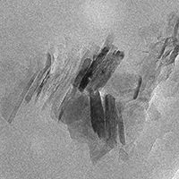
Transmission electron microscopy (TEM) characterization of MMT-Epoxy nanocomposite coatings obtained by electrophoretic deposition process
All claims expressed in this article are solely those of the authors and do not necessarily represent those of their affiliated organizations, or those of the publisher, the editors and the reviewers. Any product that may be evaluated in this article or claim that may be made by its manufacturer is not guaranteed or endorsed by the publisher.
Published: 2 March 2021
Epoxy-nanoclay composite coatings are particularly attractive for their application in many technological areas, such as anticorrosion coatings on metal substrates and protective barriers. By adding layered silicates, having typically a structure formed by platelets with regular interspaces, it is possible to achieve an improvement of the specific properties of epoxy, even with surprising results. This is due to the penetration of polymer matrix between the silicate layers, inducing a dispersion of nanoplatelets at different degrees. In order to maximize the positive effect of the added silicate on the properties of the epoxy matrix, it is of primary importance to optimize the coating preparation process. To this aim, transmission electron microscopy (TEM) is an invaluable characterization technique, essential to obtain complete information on how much and how the nanoplatelets are distributed in the polymer matrix. For TEM observations it is necessary to find the right way to prepare a section of the sample by ultramicrotomy, without introducing artifacts. In this paper, we report TEM studies of montmorillonite-epoxy (MMT-epoxy) coatings obtained by Electrophoretic Deposition (EPD).
Downloads
How to Cite
PAGEPress has chosen to apply the Creative Commons Attribution NonCommercial 4.0 International License (CC BY-NC 4.0) to all manuscripts to be published.
An Open Access Publication is one that meets the following two conditions:
- the author(s) and copyright holder(s) grant(s) to all users a free, irrevocable, worldwide, perpetual right of access to, and a license to copy, use, distribute, transmit and display the work publicly and to make and distribute derivative works, in any digital medium for any responsible purpose, subject to proper attribution of authorship, as well as the right to make small numbers of printed copies for their personal use.
- a complete version of the work and all supplemental materials, including a copy of the permission as stated above, in a suitable standard electronic format is deposited immediately upon initial publication in at least one online repository that is supported by an academic institution, scholarly society, government agency, or other well-established organization that seeks to enable open access, unrestricted distribution, interoperability, and long-term archiving.
Authors who publish with this journal agree to the following terms:
- Authors retain copyright and grant the journal right of first publication with the work simultaneously licensed under a Creative Commons Attribution License that allows others to share the work with an acknowledgement of the work's authorship and initial publication in this journal.
- Authors are able to enter into separate, additional contractual arrangements for the non-exclusive distribution of the journal's published version of the work (e.g., post it to an institutional repository or publish it in a book), with an acknowledgement of its initial publication in this journal.
- Authors are permitted and encouraged to post their work online (e.g., in institutional repositories or on their website) prior to and during the submission process, as it can lead to productive exchanges, as well as earlier and greater citation of published work.