Contribution of light and electron microscopy in the identification of morphological alterations in large Japanese field mouse (Apodemus speciosus) testes exposed to low-dose-rate radiations
All claims expressed in this article are solely those of the authors and do not necessarily represent those of their affiliated organizations, or those of the publisher, the editors and the reviewers. Any product that may be evaluated in this article or claim that may be made by its manufacturer is not guaranteed or endorsed by the publisher.
Published: 2 March 2021
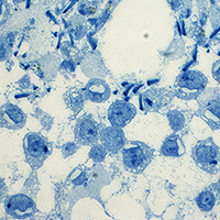
Ionizing radiation affects biological systems, resulting in an increased risk of cancer and mutagenesis. Male reproductive function is sensitive to ionizing radiation, with implications connected to infertility. Following the Nuclear Power Plants accident of Fukushima in 2011, there was great attention regarding exposure damage to low-dose-rate (LDR) radiation on the reproductive system. This preliminary study aimed to evaluate the role of light (LM) and transmission electron microscopies (TEM) to identify the potential effects of LDR radio-exposure on the morphology of large Japanese field mouse (Apodemus speciosus) testis testes living in the Fukushima Daiichi Nuclear Power Plant (FDNPP) ex-evacuation area. After collection samples were subjected to the standard preparative for LM and TEM. The testicular parenchyma was characterized by numerous seminiferous tubules, delimited by a thick and continuous basal lamina. Basally, the germinal epithelium presented round and pale spermatogonia, primary spermatocytes; while, more adluminally, round and elongated spermatids were at different phases of development. Pale and irregular Sertoli cells were interspersed among germ cells. Occasionally, cytoplasmatic holes interrupted the nuclear membrane integrity in spermatocytes and spermatids. Residual bodies were seen at the luminal surface. In conclusion, this study suggests that LM and TEM analysis are useful in evaluating potential morphological features in the male reproductive system after LDR exposure.
Downloads
How to Cite
PAGEPress has chosen to apply the Creative Commons Attribution NonCommercial 4.0 International License (CC BY-NC 4.0) to all manuscripts to be published.
An Open Access Publication is one that meets the following two conditions:
- the author(s) and copyright holder(s) grant(s) to all users a free, irrevocable, worldwide, perpetual right of access to, and a license to copy, use, distribute, transmit and display the work publicly and to make and distribute derivative works, in any digital medium for any responsible purpose, subject to proper attribution of authorship, as well as the right to make small numbers of printed copies for their personal use.
- a complete version of the work and all supplemental materials, including a copy of the permission as stated above, in a suitable standard electronic format is deposited immediately upon initial publication in at least one online repository that is supported by an academic institution, scholarly society, government agency, or other well-established organization that seeks to enable open access, unrestricted distribution, interoperability, and long-term archiving.
Authors who publish with this journal agree to the following terms:
- Authors retain copyright and grant the journal right of first publication with the work simultaneously licensed under a Creative Commons Attribution License that allows others to share the work with an acknowledgement of the work's authorship and initial publication in this journal.
- Authors are able to enter into separate, additional contractual arrangements for the non-exclusive distribution of the journal's published version of the work (e.g., post it to an institutional repository or publish it in a book), with an acknowledgement of its initial publication in this journal.
- Authors are permitted and encouraged to post their work online (e.g., in institutional repositories or on their website) prior to and during the submission process, as it can lead to productive exchanges, as well as earlier and greater citation of published work.