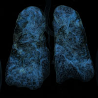
Overview of COVID-19 patients treated in University Hospital Split, Croatia - specifics related to patients age

Accepted: 14 January 2021
HTML: 9
All claims expressed in this article are solely those of the authors and do not necessarily represent those of their affiliated organizations, or those of the publisher, the editors and the reviewers. Any product that may be evaluated in this article or claim that may be made by its manufacturer is not guaranteed or endorsed by the publisher.
Different aspects of the coronavirus disease 2019 (COVID-19) infection have been widely investigated since the onset of a pandemic in December 2019. Several studies investigated differences in disease development and presentation compared to patient characteristics. In this paper we present an overview of the first COVID-19 pandemic wave in Dalmatia, Croatia with specifics related to patients’ age. Demographic, clinical and radiological data from hospitalized COVID-19 positive patients in the Clinical Hospital Split over a three-month period were collected and analyzed. Subgrouping and additional analysis were performed: Octogenarians vs Non-octogenarians, and senior residence vs nonsenior residence. 160 COVID-19 positive patients were enrolled. Of those, 61% were females. Median age was 78. More than a half of all patients were senior residents. No differences in final outcome were observed comparing specific medicament treatment. Among Octogenarians group, there were more asymptomatic cases, and higher mortality rate. Some differences in radiological presentation were also observed. Senior COVID-19 positive patients are more often asymptomatic but with higher mortality rates. More attention should be paid to early detection on COVID-19 infection in the senior population.
Lu H, Stratton CW, Tang YW. Outbreak of pneumonia of unknown etiology in Wuhan, China: The mystery and the miracle. J Med Virol 2020;92:401-2. DOI: https://doi.org/10.1002/jmv.25678
World Health Organization (WHO). Weekly operational update on COVID-19 - 7 December 2020. Available from: https://www.who.int/publications/m/item/weekly-operational-update-on-covid-19---7-december-2020
Dennie C, Hague C, Lim RS, et al. Canadian Society of Thoracic Radiology/Canadian Association of Radiologists Consensus Statement Regarding Chest Imaging in Suspected and Confirmed COVID-19. Canad Assoc Radiol J 2020 May 8:846537120924606. DOI: https://doi.org/10.1177/0846537120924606
Wong HYF, Lam HYS, Fong AH, et al. Frequency and distribution of chest radiographic findings in patients positive for COVID-19. Radiology 2020;296:E72-E8. DOI: https://doi.org/10.1148/radiol.2020201160
World Health Organization (WHO). Clinical management of COVID-19; 27 May 2020. Available from: https://www.who.int/publications/i/item/clinical-management-of-covid-19
World Health Organization (WHO). Life tables by country – Croatia; Last updated 6 December 2020. Available from: https://apps.who.int/gho/data/?theme=main&vid=60400
Oran DP, Topol EJ. Prevalence of asymptomatic SARS-CoV-2 infection: a narrative review. Ann Intern Med 2020 [Epub ahead of print]. DOI: https://doi.org/10.7326/M20-3012
Li LQ, Huang T, Wang YQ, et al. COVID-19 patients’ clinical characteristics, discharge rate, and fatality rate of meta-analysis. J Med Virol 2020;92:577-83. DOI: https://doi.org/10.1002/jmv.25757
Cheung KS, Hung IFN, Chan PPY, et al. Gastrointestinal manifestations of SARS-CoV-2 infection and virus load in fecal samples from a Hong Kong cohort: systematic review and meta-analysis. Gastroenterology 2020;159:81-95. DOI: https://doi.org/10.1053/j.gastro.2020.03.065
Jin X, Lian JS, Hu JH, et al. Epidemiological, clinical and virological characteristics of 74 cases of coronavirus-infected disease 2019 (COVID-19) with gastrointestinal symptoms. Gut 2020;69:1002-9. DOI: https://doi.org/10.1136/gutjnl-2020-320926
Uyeki TM, Holshue ML, Diaz G. First Case of Covid-19 in the United States. Reply. N Engl J Med 2020;382:e53. DOI: https://doi.org/10.1056/NEJMc2004794
Wang D, Hu B, Hu C, et al. Clinical characteristics of 138 hospitalized patients with 2019 novel coronavirus-infected pneumonia in Wuhan, China. JAMA 2020 [Epub ahead of print]. DOI: https://doi.org/10.1001/jama.2020.1585
Karimi-Galougahi M, Naeini AS, Raad N, et al. Vertigo and hearing loss during the COVID-19 pandemic - is there an association? Acta Otorhinolaryngol Italica 2020 [Epub ahead of print]. DOI: https://doi.org/10.14639/0392-100X-N0820
Suarez-Valle A, Fernandez-Nieto D, Diaz-Guimaraens B, et al. Acro-ischaemia in hospitalized COVID-19 patients. J Eur Acad Dermatol Venereol 2020 [Epub ahead of print]. DOI: https://doi.org/10.1111/jdv.16592
Adams SJ, Dennie C. Chest imaging in patients with suspected COVID-19. CMAJ Canad Med Assoc J 2020;192:E676. DOI: https://doi.org/10.1503/cmaj.200626
Bernheim A, Mei X, Huang M, et al. Chest CT findings in coronavirus disease-19 (COVID-19): relationship to duration of infection. Radiology 2020;295:200463. DOI: https://doi.org/10.1148/radiol.2020200463
Ye Z, Zhang Y, Wang Y, et al. Chest CT manifestations of new coronavirus disease 2019 (COVID-19): a pictorial review. Eur Radiol 2020;30:4381-9. DOI: https://doi.org/10.1007/s00330-020-06801-0
Sun R, Liu H, Wang X. Mediastinal emphysema, giant bulla, and pneumothorax developed during the course of COVID-19 pneumonia. Korean J Radiol 2020;21:541-4. DOI: https://doi.org/10.3348/kjr.2020.0180
Ucpinar BA, Sahin C, Yanc U. Spontaneous pneumothorax and subcutaneous emphysema in COVID-19 patient: case report. J Infect Public Health 2020;13:887-9. DOI: https://doi.org/10.1016/j.jiph.2020.05.012
Wang W, Gao R, Zheng Y, Jiang L. COVID-19 with spontaneous pneumothorax, pneumomediastinum and subcutaneous emphysema. J Travel Med 2020 [Epub ahead of print]. DOI: https://doi.org/10.1093/jtm/taaa062
Gautret P, Lagier JC, Parola P, et al. Clinical and microbiological effect of a combination of hydroxychloroquine and azithromycin in 80 COVID-19 patients with at least a six-day follow up: A pilot observational study. Travel Med Infect Dis 2020;34:101663. DOI: https://doi.org/10.1016/j.tmaid.2020.101663
Gbinigie K, Frie K. Should azithromycin be used to treat COVID-19? A rapid review. BJGP Open 2020;4:2. DOI: https://doi.org/10.3399/bjgpopen20X101094
Koo HJ, Lim S, Choe J, et al. Radiographic and CT features of viral pneumonia. Radiographics 2018;38:719-39. DOI: https://doi.org/10.1148/rg.2018170048
PAGEPress has chosen to apply the Creative Commons Attribution NonCommercial 4.0 International License (CC BY-NC 4.0) to all manuscripts to be published.


 https://doi.org/10.4081/gc.2021.9351


