
The importance of sonographic evaluation of muscle depth and thickness prior to the ‘tiny percutaneous needle biopsy’
HTML: 88
All claims expressed in this article are solely those of the authors and do not necessarily represent those of their affiliated organizations, or those of the publisher, the editors and the reviewers. Any product that may be evaluated in this article or claim that may be made by its manufacturer is not guaranteed or endorsed by the publisher.
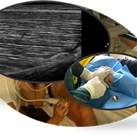
Biopsy of human skeletal muscle tissue is a widely used method in many research studies, where ‘the tiny percutaneous needle biopsy’ (TPNB) is one of the relatively simplest and safest procedures currently available. By using and contrasting ultrasound images of vastus lateralis of young and elderly subjects, this work highlights further the safety aspects of TPNB and stresses the importance of prior ultrasound evaluation of muscle depth and thickness in order to prevent wrong muscle group or tissue sampling in subsequent laboratory analyses.
Dodds RM, Davies K, Granic A, Hollingsworth KG, Warren C, Gorman G, et al. Mitochondrial respiratory chain function and content are preserved in the skeletal muscle of active very old men and women. Exp Gerontol. 2018;113:80–5. DOI: https://doi.org/10.1016/j.exger.2018.09.020
Franchi MV, Atherton PJ, Reeves ND, Flück M, Williams J, Mitchell WK, et al. Architectural, functional and molecular responses to concentric and eccentric loading in human skeletal muscle. Acta Physiol (Oxf). 2014 Mar;210(3):642–54. DOI: https://doi.org/10.1111/apha.12225
Wagner KR, Fleckenstein JL, Amato AA, Barohn RJ, Bushby K, Escolar DM, et al. A phase I/IItrial of MYO-029 in adult subjects with muscular dystrophy. Ann Neurol. 2008 May;63(5):561–71. DOI: https://doi.org/10.1002/ana.21338
Dietrichson P, Coakley J, Smith PE, Griffiths RD, Helliwell TR, Edwards RH. Conchotome and needle percutaneous biopsy of skeletal muscle. J Neurol Neurosurg Psychiatry. 1987 Nov;50(11):1461–7. DOI: https://doi.org/10.1136/jnnp.50.11.1461
Pietrangelo T, D’Amelio L, Doria C, Mancinelli R, Fulle S, Fanò G. Tiny percutaneous needle biopsy: An efficient method for studying cellular and molecular aspects of skeletal muscle in humans. Int J Mol Med. 2011 Mar;27(3):361–7. DOI: https://doi.org/10.3892/ijmm.2010.582
Pietrangelo T, Perni S, Di Tano G, Fanò-Illic G, Franzini-Armstrong C. A method for the ultrastructural preservation of tiny percutaneous needle biopsy material from skeletal muscle. Int J Mol Med. 2013 Oct;32(4):965–70. DOI: https://doi.org/10.3892/ijmm.2013.1454
O’Sullivan PJ, Gorman GM, Hardiman OM, Farrell MJ, Logan PM. Sonographically guided percutaneous muscle biopsy in diagnosis of neuromuscular disease: a useful alternative to open surgical biopsy. J Ultrasound Med. 2006 Jan;25(1):1–6. DOI: https://doi.org/10.7863/jum.2006.25.1.1
Wilson D, Breen L, Lord JM, Sapey E. The challenges of muscle biopsy in a community based geriatric population. BMC Res Notes. 2018 Nov 26;11(1):830. DOI: https://doi.org/10.1186/s13104-018-3947-8
Ryall JG, Schertzer JD, Lynch GS. Cellular and molecular mechanisms underlying age-related skeletal muscle wasting and weakness. Biogerontology. 2008 Aug;9(4):213–28. DOI: https://doi.org/10.1007/s10522-008-9131-0
Welle S. Cellular and molecular basis of age-related sarcopenia. Can J Appl Physiol. 2002 Feb;27(1):19–41. DOI: https://doi.org/10.1139/h02-002
Verdijk LB, Dirks ML, Snijders T, Prompers JJ, Beelen M, Jonkers RAM, et al. Reduced satellite cell numbers with spinal cord injury and aging in humans. Med Sci Sports Exerc. 2012 Dec;44(12):2322–30. DOI: https://doi.org/10.1249/MSS.0b013e3182667c2e
Ticinesi A, Narici MV, Lauretani F, Nouvenne A, Colizzi E, Mantovani M, et al. Assessing sarcopenia with vastus lateralis muscle ultrasound: an operative protocol. Aging Clin Exp Res. 2018 Dec;30(12):1437–43. DOI: https://doi.org/10.1007/s40520-018-0958-1
Kemp GJ, Birrell F, Clegg PD, Cuthbertson DJ, De Vito G, van Dieën JH, et al. Developing a toolkit for the assessment and monitoring of musculoskeletal ageing. Age and Ageing. 2018 Sep 1;47(suppl_4):iv1–19. DOI: https://doi.org/10.1093/ageing/afy143
PAGEPress has chosen to apply the Creative Commons Attribution NonCommercial 4.0 International License (CC BY-NC 4.0) to all manuscripts to be published.
Downloads
Citations


